 Movie
Movie Controller


+ Open data

- Basic information
| Entry | Database: PDB / ID: 6w4s | |||||||||||||||||||||||||||||||||
|---|---|---|---|---|---|---|---|---|---|---|---|---|---|---|---|---|---|---|---|---|---|---|---|---|---|---|---|---|---|---|---|---|---|---|
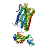
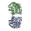
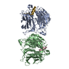
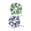
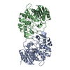
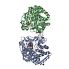
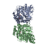
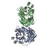
| Title | Structure of apo human ferroportin in lipid nanodisc | |||||||||||||||||||||||||||||||||
 Components Components |
| |||||||||||||||||||||||||||||||||
 Keywords Keywords | TRANSPORT PROTEIN/IMMUNE SYSTEM / ferroportin / transporter / iron / hepcidin / TRANSPORT PROTEIN-IMMUNE SYSTEM complex | |||||||||||||||||||||||||||||||||
| Function / homology |  Function and homology information Function and homology informationspleen trabecula formation / iron ion export across plasma membrane / Defective SLC40A1 causes hemochromatosis 4 (HFE4) (duodenum) / Defective SLC40A1 causes hemochromatosis 4 (HFE4) (macrophages) / Defective CP causes aceruloplasminemia (ACERULOP) / Metal ion SLC transporters / lymphocyte homeostasis / ferrous iron transmembrane transporter activity / iron ion transmembrane transporter activity / endothelium development ...spleen trabecula formation / iron ion export across plasma membrane / Defective SLC40A1 causes hemochromatosis 4 (HFE4) (duodenum) / Defective SLC40A1 causes hemochromatosis 4 (HFE4) (macrophages) / Defective CP causes aceruloplasminemia (ACERULOP) / Metal ion SLC transporters / lymphocyte homeostasis / ferrous iron transmembrane transporter activity / iron ion transmembrane transporter activity / endothelium development / iron ion transmembrane transport / peptide hormone binding / establishment of localization in cell / Iron uptake and transport / multicellular organismal-level iron ion homeostasis / synaptic vesicle / transcription by RNA polymerase II / basolateral plasma membrane / intracellular iron ion homeostasis / apoptotic process / negative regulation of apoptotic process / positive regulation of transcription by RNA polymerase II / nucleoplasm / membrane / metal ion binding / identical protein binding / plasma membrane / cytosol / cytoplasm Similarity search - Function | |||||||||||||||||||||||||||||||||
| Biological species |  Homo sapiens (human) Homo sapiens (human) | |||||||||||||||||||||||||||||||||
| Method | ELECTRON MICROSCOPY / single particle reconstruction / cryo EM / Resolution: 3.2 Å | |||||||||||||||||||||||||||||||||
 Authors Authors | Billesboelle, C.B. / Azumaya, C.M. / Gonen, S. / Powers, A. / Kretsch, R.C. / Schneider, S. / Arvedson, T. / Dror, R.O. / Cheng, Y. / Manglik, A. | |||||||||||||||||||||||||||||||||
| Funding support |  United States, 1items United States, 1items
| |||||||||||||||||||||||||||||||||
 Citation Citation | Journal: Nature / Year: 2020 Title: Structure of hepcidin-bound ferroportin reveals iron homeostatic mechanisms. Authors: Christian B Billesbølle / Caleigh M Azumaya / Rachael C Kretsch / Alexander S Powers / Shane Gonen / Simon Schneider / Tara Arvedson / Ron O Dror / Yifan Cheng / Aashish Manglik /  Abstract: The serum level of iron in humans is tightly controlled by the action of the hormone hepcidin on the iron efflux transporter ferroportin. Hepcidin regulates iron absorption and recycling by inducing ...The serum level of iron in humans is tightly controlled by the action of the hormone hepcidin on the iron efflux transporter ferroportin. Hepcidin regulates iron absorption and recycling by inducing the internalization and degradation of ferroportin. Aberrant ferroportin activity can lead to diseases of iron overload, such as haemochromatosis, or iron limitation anaemias. Here we determine cryogenic electron microscopy structures of ferroportin in lipid nanodiscs, both in the apo state and in complex with hepcidin and the iron mimetic cobalt. These structures and accompanying molecular dynamics simulations identify two metal-binding sites within the N and C domains of ferroportin. Hepcidin binds ferroportin in an outward-open conformation and completely occludes the iron efflux pathway to inhibit transport. The carboxy terminus of hepcidin directly contacts the divalent metal in the ferroportin C domain. Hepcidin binding to ferroportin is coupled to iron binding, with an 80-fold increase in hepcidin affinity in the presence of iron. These results suggest a model for hepcidin regulation of ferroportin, in which only ferroportin molecules loaded with iron are targeted for degradation. More broadly, our structural and functional insights may enable more targeted manipulation of the hepcidin-ferroportin axis in disorders of iron homeostasis. | |||||||||||||||||||||||||||||||||
| History |
|
- Structure visualization
| Movie |
Movie viewer |
|---|---|
| Structure viewer | Molecule: MolmilJmol/JSmol |
- Downloads & links
-Download
| PDBx/mmCIF format | 6w4s.cif.gz | 131.1 KB | Display | PDBx/mmCIF format |
|---|---|---|---|---|
| PDB format | pdb6w4s.ent.gz | 95.3 KB | Display | PDB format |
| PDBx/mmJSON format | 6w4s.json.gz | Tree view | PDBx/mmJSON format | |
| Others |  Other downloads Other downloads |
-Validation report
| Arichive directory | https://data.pdbj.org/pub/pdb/validation_reports/w4/6w4sftp://data.pdbj.org/pub/pdb/validation_reports/w4/6w4s | HTTPS FTP |
|---|
-Related structure data
| Related structure data |  21539MC  6w4vC  6wbvC M: map data used to model this data C: citing same article ( |
|---|---|
| Similar structure data |


-Links
 PDBj
PDBj


- Assembly
| Deposited unit | 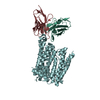
|
|---|---|
| 1 |
|
-Components
| #1: Protein | Mass: 66349.898 Da / Num. of mol.: 1 Source method: isolated from a genetically manipulated source Source: (gene. exp.) Homo sapiens (human) / Gene: SLC40A1, FPN1, IREG1, SLC11A3, MSTP079 / Production host:   Spodoptera frugiperda (fall armyworm) / References: UniProt: Q9NP59 Spodoptera frugiperda (fall armyworm) / References: UniProt: Q9NP59 |
|---|---|
| #2: Protein | Mass: 23852.592 Da / Num. of mol.: 1 Source method: isolated from a genetically manipulated source Source: (gene. exp.) Homo sapiens (human) |
| #3: Antibody | Mass: 24008.516 Da / Num. of mol.: 1 Source method: isolated from a genetically manipulated source Source: (gene. exp.) Homo sapiens (human) |
| Has protein modification | Y |
-Experimental details
-Experiment
| Experiment | Method: ELECTRON MICROSCOPY |
|---|---|
| EM experiment | Aggregation state: PARTICLE / 3D reconstruction method: single particle reconstruction |
- Sample preparation
| Component |
| ||||||||||||||||||||||||
|---|---|---|---|---|---|---|---|---|---|---|---|---|---|---|---|---|---|---|---|---|---|---|---|---|---|
| Molecular weight | Experimental value: NO | ||||||||||||||||||||||||
| Source (natural) |
| ||||||||||||||||||||||||
| Source (recombinant) |
| ||||||||||||||||||||||||
| Buffer solution | pH: 7.5 | ||||||||||||||||||||||||
| Specimen | Embedding applied: NO / Shadowing applied: NO / Staining applied: NO / Vitrification applied: YES | ||||||||||||||||||||||||
| Specimen support | Details: unspecified | ||||||||||||||||||||||||
| Vitrification | Cryogen name: ETHANE |
- Electron microscopy imaging
| Experimental equipment |  Model: Titan Krios / Image courtesy: FEI Company |
|---|---|
| Microscopy | Model: FEI TITAN KRIOS |
| Electron gun | Electron source:  FIELD EMISSION GUN / Accelerating voltage: 300 kV / Illumination mode: FLOOD BEAM FIELD EMISSION GUN / Accelerating voltage: 300 kV / Illumination mode: FLOOD BEAM |
| Electron lens | Mode: BRIGHT FIELD / Nominal magnification: 105000 X / Nominal defocus max: -20000 nm / Nominal defocus min: -8000 nm / Cs: 2.7 mm / C2 aperture diameter: 70 µm / Alignment procedure: COMA FREE |
| Specimen holder | Cryogen: NITROGEN / Specimen holder model: FEI TITAN KRIOS AUTOGRID HOLDER |
| Image recording | Average exposure time: 6 sec. / Electron dose: 60 e/Å2 / Film or detector model: GATAN K3 (6k x 4k) / Num. of grids imaged: 3 / Num. of real images: 5415 |
| EM imaging optics | Energyfilter slit width: 20 eV |
- Processing
| Software | Name: PHENIX / Version: 1.17.1_3660: / Classification: refinement | |||||||||||||||||||||||||||
|---|---|---|---|---|---|---|---|---|---|---|---|---|---|---|---|---|---|---|---|---|---|---|---|---|---|---|---|---|
| EM software |
| |||||||||||||||||||||||||||
| CTF correction | Type: PHASE FLIPPING AND AMPLITUDE CORRECTION | |||||||||||||||||||||||||||
| Symmetry | Point symmetry: C1 (asymmetric) | |||||||||||||||||||||||||||
| 3D reconstruction | Resolution: 3.2 Å / Resolution method: FSC 0.143 CUT-OFF / Num. of particles: 308366 / Symmetry type: POINT | |||||||||||||||||||||||||||
| Refine LS restraints |
|
