Movie
Movie Controller

[English] 日本語
 Yorodumi
- PDB-6w17: Structure of Dip1-activated Arp2/3 complex with nucleated actin f... -
+ Open data

- Basic information
| Entry | Database: PDB / ID: 6w17 | ||||||||||||||||||||||||||||||||||||||||||||||||||||||||||||||||||||||||||||||||||||||||||||||||
|---|---|---|---|---|---|---|---|---|---|---|---|---|---|---|---|---|---|---|---|---|---|---|---|---|---|---|---|---|---|---|---|---|---|---|---|---|---|---|---|---|---|---|---|---|---|---|---|---|---|---|---|---|---|---|---|---|---|---|---|---|---|---|---|---|---|---|---|---|---|---|---|---|---|---|---|---|---|---|---|---|---|---|---|---|---|---|---|---|---|---|---|---|---|---|---|---|---|
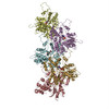
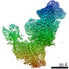
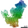
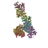
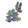
| Title | Structure of Dip1-activated Arp2/3 complex with nucleated actin filament | ||||||||||||||||||||||||||||||||||||||||||||||||||||||||||||||||||||||||||||||||||||||||||||||||
 Components Components |
| ||||||||||||||||||||||||||||||||||||||||||||||||||||||||||||||||||||||||||||||||||||||||||||||||
 Keywords Keywords | STRUCTURAL PROTEIN / Arp2/3 / actin / Dip1 / cytoskeletal protein / actin regulator | ||||||||||||||||||||||||||||||||||||||||||||||||||||||||||||||||||||||||||||||||||||||||||||||||
| Function / homology |  Function and homology information Function and homology informationprotein localization to actin cortical patch / Regulation of actin dynamics for phagocytic cup formation / RHO GTPases Activate WASPs and WAVEs / Clathrin-mediated endocytosis / actin cortical patch organization / medial cortex / Neutrophil degranulation / actin filament branching / cell cortex of cell tip / actin cortical patch localization ...protein localization to actin cortical patch / Regulation of actin dynamics for phagocytic cup formation / RHO GTPases Activate WASPs and WAVEs / Clathrin-mediated endocytosis / actin cortical patch organization / medial cortex / Neutrophil degranulation / actin filament branching / cell cortex of cell tip / actin cortical patch localization / actin cortical patch assembly / Arp2/3 protein complex / Arp2/3 complex-mediated actin nucleation / Arp2/3 complex binding / actin cortical patch / cell tip / regulation of actin filament polymerization / mating projection tip / cortical actin cytoskeleton organization / cytoskeletal motor activator activity / cell division site / myosin heavy chain binding / tropomyosin binding / actin filament bundle / troponin I binding / filamentous actin / mesenchyme migration / cortical cytoskeleton / establishment or maintenance of cell polarity / skeletal muscle myofibril / actin filament bundle assembly / striated muscle thin filament / skeletal muscle thin filament assembly / actin monomer binding / skeletal muscle fiber development / stress fiber / titin binding / actin filament polymerization / filopodium / actin filament / structural constituent of cytoskeleton / Hydrolases; Acting on acid anhydrides; Acting on acid anhydrides to facilitate cellular and subcellular movement / endocytosis / calcium-dependent protein binding / actin filament binding / lamellipodium / toxin activity / cell body / cell cortex / protein-macromolecule adaptor activity / protein domain specific binding / hydrolase activity / calcium ion binding / positive regulation of gene expression / magnesium ion binding / ATP binding / identical protein binding / nucleus / cytoplasm / cytosol Similarity search - Function | ||||||||||||||||||||||||||||||||||||||||||||||||||||||||||||||||||||||||||||||||||||||||||||||||
| Biological species |    Amanita phalloides (death cap) Amanita phalloides (death cap) | ||||||||||||||||||||||||||||||||||||||||||||||||||||||||||||||||||||||||||||||||||||||||||||||||
| Method | ELECTRON MICROSCOPY / single particle reconstruction / cryo EM / Resolution: 3.9 Å | ||||||||||||||||||||||||||||||||||||||||||||||||||||||||||||||||||||||||||||||||||||||||||||||||
 Authors Authors | Shaaban, M. / Nolen, B.J. / Chowdhury, S. | ||||||||||||||||||||||||||||||||||||||||||||||||||||||||||||||||||||||||||||||||||||||||||||||||
| Funding support |  United States, 1items United States, 1items
| ||||||||||||||||||||||||||||||||||||||||||||||||||||||||||||||||||||||||||||||||||||||||||||||||
 Citation Citation | Journal: Nat Struct Mol Biol / Year: 2020 Title: Cryo-EM reveals the transition of Arp2/3 complex from inactive to nucleation-competent state. Authors: Mohammed Shaaban / Saikat Chowdhury / Brad J Nolen / Abstract: Arp2/3 complex, a crucial actin filament nucleator, undergoes structural rearrangements during activation by nucleation-promoting factors (NPFs). However, the conformational pathway leading to the ...Arp2/3 complex, a crucial actin filament nucleator, undergoes structural rearrangements during activation by nucleation-promoting factors (NPFs). However, the conformational pathway leading to the nucleation-competent state is unclear due to lack of high-resolution structures of the activated state. Here we report a ~3.9 Å resolution cryo-EM structure of activated Schizosaccharomyces pombe Arp2/3 complex bound to the S. pombe NPF Dip1 and attached to the end of the nucleated actin filament. The structure reveals global and local conformational changes that allow the two actin-related proteins in Arp2/3 complex to mimic a filamentous actin dimer and template nucleation. Activation occurs through a clamp-twisting mechanism, in which Dip1 forces two core subunits in Arp2/3 complex to pivot around one another, shifting half of the complex into a new activated position. By showing how Dip1 stimulates activation, the structure reveals how NPFs can activate Arp2/3 complex in diverse cellular processes. | ||||||||||||||||||||||||||||||||||||||||||||||||||||||||||||||||||||||||||||||||||||||||||||||||
| History |
|
- Structure visualization
| Movie |
Movie viewer |
|---|---|
| Structure viewer | Molecule: MolmilJmol/JSmol |
- Downloads & links
-Download
| PDBx/mmCIF format | 6w17.cif.gz | 634.2 KB | Display | PDBx/mmCIF format |
|---|---|---|---|---|
| PDB format | pdb6w17.ent.gz | 500.1 KB | Display | PDB format |
| PDBx/mmJSON format | 6w17.json.gz | Tree view | PDBx/mmJSON format | |
| Others |  Other downloads Other downloads |
-Validation report
| Arichive directory | https://data.pdbj.org/pub/pdb/validation_reports/w1/6w17ftp://data.pdbj.org/pub/pdb/validation_reports/w1/6w17 | HTTPS FTP |
|---|
-Related structure data
| Related structure data | 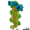 21502MC  6w18C M: map data used to model this data C: citing same article ( |
|---|---|
| Similar structure data |


-Links
 PDBj
PDBj


- Assembly
| Deposited unit | 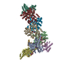
|
|---|---|
| 1 |
|
-Components
-Actin-related protein ... , 7 types, 7 molecules ABCDEFG
| #1: Protein | Mass: 47427.137 Da / Num. of mol.: 1 / Source method: isolated from a natural source Source: (natural) Plasmid details: Fission yeast / Strain: 972 / ATCC 24843 / References: UniProt: P32390 |
|---|---|
| #2: Protein | Mass: 44286.758 Da / Num. of mol.: 1 / Source method: isolated from a natural source Source: (natural) Plasmid details: Fission yeast / Strain: 972 / ATCC 24843 / References: UniProt: Q9UUJ1 |
| #3: Protein | Mass: 41643.465 Da / Num. of mol.: 1 / Source method: isolated from a natural source Source: (natural) Plasmid details: Fission yeast / Strain: 972 / ATCC 24843 / References: UniProt: P78774 |
| #4: Protein | Mass: 37025.230 Da / Num. of mol.: 1 / Source method: isolated from a natural source Source: (natural) Plasmid details: Fission yeast / Strain: 972 / ATCC 24843 / References: UniProt: O14241 |
| #5: Protein | Mass: 19865.746 Da / Num. of mol.: 1 / Source method: isolated from a natural source Source: (natural) Plasmid details: Fission yeast / Strain: 972 / ATCC 24843 / References: UniProt: Q9Y7J4 |
| #6: Protein | Mass: 19637.695 Da / Num. of mol.: 1 / Source method: isolated from a natural source Source: (natural) Plasmid details: Fission yeast / Strain: 972 / ATCC 24843 / References: UniProt: Q92352 |
| #7: Protein | Mass: 16922.059 Da / Num. of mol.: 1 / Source method: isolated from a natural source Source: (natural) Plasmid details: Fission yeast / Strain: 972 / ATCC 24843 / References: UniProt: Q10316 |
-Protein , 2 types, 5 molecules HIJKL
| #8: Protein | Mass: 43715.137 Da / Num. of mol.: 1 Source method: isolated from a genetically manipulated source Details: Fission yeast Source: (gene. exp.) Strain: 972 / ATCC 24843 / Gene: dip1, SPBC24C6.10c / Plasmid: pGV67-Dip1 / Production host:  |
|---|---|
| #9: Protein | Mass: 42109.973 Da / Num. of mol.: 4 / Source method: isolated from a natural source / Source: (natural) |
-Protein/peptide , 1 types, 5 molecules MNOPQ
| #10: Protein/peptide | Mass: 808.899 Da / Num. of mol.: 5 / Source method: isolated from a natural source / Source: (natural) Amanita phalloides (death cap) / References: UniProt: P0CU63*PLUS |
|---|
-Non-polymers , 3 types, 12 molecules 


| #11: Chemical | ChemComp-MG /  Mass: 24.305 Da / Num. of mol.: 6 / Source method: obtained synthetically / Formula: Mg Mass: 24.305 Da / Num. of mol.: 6 / Source method: obtained synthetically / Formula: Mg#12: Chemical |  Mass: 507.181 Da / Num. of mol.: 2 / Source method: obtained synthetically / Formula: C10H16N5O13P3 / Comment: ATP, energy-carrying molecule*YM Mass: 507.181 Da / Num. of mol.: 2 / Source method: obtained synthetically / Formula: C10H16N5O13P3 / Comment: ATP, energy-carrying molecule*YM#13: Chemical | ChemComp-ADP /  Mass: 427.201 Da / Num. of mol.: 4 / Source method: obtained synthetically / Formula: C10H15N5O10P2 / Comment: ADP, energy-carrying molecule*YM Mass: 427.201 Da / Num. of mol.: 4 / Source method: obtained synthetically / Formula: C10H15N5O10P2 / Comment: ADP, energy-carrying molecule*YM |
|---|
-Details
| Has ligand of interest | N |
|---|---|
| Has protein modification | Y |
-Experimental details
-Experiment
| Experiment | Method: ELECTRON MICROSCOPY |
|---|---|
| EM experiment | Aggregation state: PARTICLE / 3D reconstruction method: single particle reconstruction |
- Sample preparation
| Component | Name: Complex consisting of actin-filament nucleator, Arp2/3 complex associated with nucleation promoting factor Dip1 and first four actin subunits from the pointed end of the nucleated actin-filament Type: COMPLEX / Entity ID: #1-#10 / Source: NATURAL | ||||||||||||||||||||||||||||||||||||||||
|---|---|---|---|---|---|---|---|---|---|---|---|---|---|---|---|---|---|---|---|---|---|---|---|---|---|---|---|---|---|---|---|---|---|---|---|---|---|---|---|---|---|
| Molecular weight | Value: 0.446 MDa / Experimental value: NO | ||||||||||||||||||||||||||||||||||||||||
| Source (natural) | Organism: Strain: 972 / ATCC 24843 | ||||||||||||||||||||||||||||||||||||||||
| Buffer solution | pH: 7.5 | ||||||||||||||||||||||||||||||||||||||||
| Buffer component |
| ||||||||||||||||||||||||||||||||||||||||
| Specimen | Conc.: 3 mg/ml / Embedding applied: NO / Shadowing applied: NO / Staining applied: NO / Vitrification applied: YES | ||||||||||||||||||||||||||||||||||||||||
| Specimen support | Details: 20mA current / Grid material: GOLD / Grid mesh size: 300 divisions/in. / Grid type: Quantifoil, UltrAuFoil, R1.2/1.3 | ||||||||||||||||||||||||||||||||||||||||
| Vitrification | Instrument: HOMEMADE PLUNGER / Cryogen name: ETHANE / Humidity: 98 % / Chamber temperature: 277.15 K |
- Electron microscopy imaging
| Experimental equipment |  Model: Talos Arctica / Image courtesy: FEI Company |
|---|---|
| Microscopy | Model: FEI TALOS ARCTICA Details: Data were collected by stage shifting to targeted exposure positions. |
| Electron gun | Electron source:  FIELD EMISSION GUN / Accelerating voltage: 200 kV / Illumination mode: FLOOD BEAM FIELD EMISSION GUN / Accelerating voltage: 200 kV / Illumination mode: FLOOD BEAM |
| Electron lens | Mode: BRIGHT FIELD / Nominal magnification: 92000 X / Nominal defocus max: -1750 nm / Nominal defocus min: -1000 nm / Cs: 2.7 mm / C2 aperture diameter: 70 µm / Alignment procedure: BASIC |
| Specimen holder | Cryogen: NITROGEN / Specimen holder model: FEI TITAN KRIOS AUTOGRID HOLDER |
| Image recording | Average exposure time: 60 sec. / Electron dose: 36.35 e/Å2 / Detector mode: COUNTING / Film or detector model: FEI FALCON III (4k x 4k) / Num. of grids imaged: 2 / Num. of real images: 5109 Details: Each micrograph was collected as dose-fractionated movies consisting of 45 fractions per movie. |
| Image scans | Sampling size: 14 µm / Width: 4000 / Height: 4000 |
- Processing
| EM software |
| |||||||||||||||||||||||||||||||||||||||||||||||||||||||
|---|---|---|---|---|---|---|---|---|---|---|---|---|---|---|---|---|---|---|---|---|---|---|---|---|---|---|---|---|---|---|---|---|---|---|---|---|---|---|---|---|---|---|---|---|---|---|---|---|---|---|---|---|---|---|---|---|
| CTF correction | Details: Particles were CTF corrected during projection matching and back projection. Type: PHASE FLIPPING AND AMPLITUDE CORRECTION | |||||||||||||||||||||||||||||||||||||||||||||||||||||||
| Particle selection | Num. of particles selected: 3500000 Details: Particles were picked using Laplacian Gaussian auto-picking. | |||||||||||||||||||||||||||||||||||||||||||||||||||||||
| Symmetry | Point symmetry: C1 (asymmetric) | |||||||||||||||||||||||||||||||||||||||||||||||||||||||
| 3D reconstruction | Resolution: 3.9 Å / Resolution method: FSC 0.143 CUT-OFF / Num. of particles: 110433 / Algorithm: BACK PROJECTION / Num. of class averages: 1 / Symmetry type: POINT | |||||||||||||||||||||||||||||||||||||||||||||||||||||||
| Atomic model building | Protocol: FLEXIBLE FIT / Space: REAL | |||||||||||||||||||||||||||||||||||||||||||||||||||||||
| Atomic model building | 3D fitting-ID: 1 / Source name: PDB / Type: experimental model
| |||||||||||||||||||||||||||||||||||||||||||||||||||||||
| Refinement | Stereochemistry target values: GeoStd + Monomer Library + CDL v1.2 | |||||||||||||||||||||||||||||||||||||||||||||||||||||||
| Displacement parameters | Biso mean: 102.46 Å2 | |||||||||||||||||||||||||||||||||||||||||||||||||||||||
| Refine LS restraints |
|