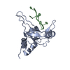
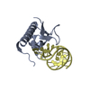
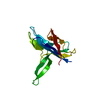
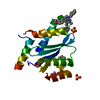
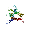
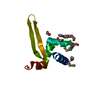
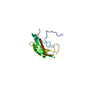
[English] 日本語
Yorodumi
- PDB-6vo7: Crystal structure of PI3K-alpha Ras Binding Domain (RBD) -

+
Open data


ID or keywords:

Loading...

-
Basic information

Entry
Database: PDB / ID: 6vo7
TitleCrystal structure of PI3K-alpha Ras Binding Domain (RBD)
ComponentsPhosphatidylinositol 4,5-bisphosphate 3-kinase catalytic subunit alpha isoform
KeywordsONCOPROTEIN / Ras binding domain / oncogenic signaling / effector
Function / homology
Function and homology information


response to muscle inactivity / negative regulation of actin filament depolymerization / response to L-leucine / regulation of actin filament organization / response to butyrate / autosome genomic imprinting / IRS-mediated signalling / cellular response to hydrostatic pressure / PI3K events in ERBB4 signaling / Activated NTRK2 signals through PI3K ...response to muscle inactivity / negative regulation of actin filament depolymerization / response to L-leucine / regulation of actin filament organization / response to butyrate / autosome genomic imprinting / IRS-mediated signalling / cellular response to hydrostatic pressure / PI3K events in ERBB4 signaling / Activated NTRK2 signals through PI3K / positive regulation of protein localization to membrane / Activated NTRK3 signals through PI3K / negative regulation of fibroblast apoptotic process / cardiac muscle cell contraction / phosphatidylinositol 3-kinase complex, class IB / vasculature development / Signaling by cytosolic FGFR1 fusion mutants / regulation of cellular respiration / phosphatidylinositol 3-kinase complex, class IA / anoikis / phosphatidylinositol 3-kinase complex / Nephrin family interactions / Costimulation by the CD28 family / relaxation of cardiac muscle / 1-phosphatidylinositol-4-phosphate 3-kinase activity / 1-phosphatidylinositol-4,5-bisphosphate 3-kinase activity / MET activates PI3K/AKT signaling / PI3K/AKT activation / phosphatidylinositol-4,5-bisphosphate 3-kinase / vascular endothelial growth factor signaling pathway / phosphatidylinositol 3-kinase / phosphatidylinositol-3-phosphate biosynthetic process / 1-phosphatidylinositol-3-kinase activity / Signaling by ALK / negative regulation of macroautophagy / PI-3K cascade:FGFR3 / Erythropoietin activates Phosphoinositide-3-kinase (PI3K) / PI-3K cascade:FGFR2 / response to dexamethasone / PI-3K cascade:FGFR4 / protein kinase activator activity / PI-3K cascade:FGFR1 / CD28 dependent PI3K/Akt signaling / Synthesis of PIPs at the plasma membrane / phosphatidylinositol-mediated signaling / phosphatidylinositol phosphate biosynthetic process / PI3K events in ERBB2 signaling / negative regulation of anoikis / RET signaling / intercalated disc / regulation of multicellular organism growth / insulin receptor substrate binding / Interleukin-3, Interleukin-5 and GM-CSF signaling / PI3K Cascade / positive regulation of TOR signaling / endothelial cell migration / RAC2 GTPase cycle / Role of phospholipids in phagocytosis / GAB1 signalosome / Role of LAT2/NTAL/LAB on calcium mobilization / Interleukin receptor SHC signaling / adipose tissue development / phagocytosis / Signaling by PDGFRA transmembrane, juxtamembrane and kinase domain mutants / Signaling by PDGFRA extracellular domain mutants / energy homeostasis / positive regulation of lamellipodium assembly / Signaling by FGFR4 in disease / cardiac muscle contraction / Signaling by FLT3 ITD and TKD mutants / Signaling by FGFR3 in disease / Tie2 Signaling / GPVI-mediated activation cascade / Signaling by FGFR2 in disease / T cell costimulation / RAC1 GTPase cycle / response to muscle stretch / Signaling by FLT3 fusion proteins / FLT3 Signaling / Signaling by FGFR1 in disease / Downstream signal transduction / phosphatidylinositol 3-kinase/protein kinase B signal transduction / insulin-like growth factor receptor signaling pathway / Signaling by phosphorylated juxtamembrane, extracellular and kinase domain KIT mutants / liver development / response to activity / Regulation of signaling by CBL / cellular response to glucose stimulus / positive regulation of smooth muscle cell proliferation / regulation of protein phosphorylation / Constitutive Signaling by EGFRvIII / Signaling by ERBB2 ECD mutants / Signaling by ERBB2 KD Mutants / epidermal growth factor receptor signaling pathway / Signaling by SCF-KIT / platelet activation / VEGFA-VEGFR2 Pathway / cellular response to insulin stimulus / Constitutive Signaling by Aberrant PI3K in Cancer / glucose metabolic process
Similarity search - Function
PI3Kalpha, catalytic domain / PI3-kinase family, p85-binding domain / PI3-kinase family, p85-binding domain / Phosphatidylinositol 3-kinase, adaptor-binding domain / Phosphatidylinositol 3-kinase adaptor-binding (PI3K ABD) domain profile. / PI3-kinase family, Ras-binding domain / Phosphatidylinositol 3-kinase Ras-binding (PI3K RBD) domain / PI3-kinase family, ras-binding domain / Phosphatidylinositol 3-kinase Ras-binding (PI3K RBD) domain profile. / Phosphoinositide 3-kinase C2 ...PI3Kalpha, catalytic domain / PI3-kinase family, p85-binding domain / PI3-kinase family, p85-binding domain / Phosphatidylinositol 3-kinase, adaptor-binding domain / Phosphatidylinositol 3-kinase adaptor-binding (PI3K ABD) domain profile. / PI3-kinase family, Ras-binding domain / Phosphatidylinositol 3-kinase Ras-binding (PI3K RBD) domain / PI3-kinase family, ras-binding domain / Phosphatidylinositol 3-kinase Ras-binding (PI3K RBD) domain profile. / Phosphoinositide 3-kinase C2 / Phosphoinositide 3-kinase, region postulated to contain C2 domain / C2 phosphatidylinositol 3-kinase-type domain / C2 phosphatidylinositol 3-kinase (PI3K)-type domain profile. / Phosphoinositide 3-kinase, accessory (PIK) domain superfamily / Phosphoinositide 3-kinase family, accessory domain (PIK domain) / Phosphoinositide 3-kinase family, accessory domain (PIK domain) / Phosphoinositide 3-kinase, accessory (PIK) domain / Phosphatidylinositol kinase / PIK helical domain profile. / Phosphatidylinositol 3- and 4-kinases signature 1. / Phosphatidylinositol 3/4-kinase, conserved site / Phosphatidylinositol 3- and 4-kinases signature 2. / Phosphatidylinositol 3-/4-kinase, catalytic domain superfamily / Phosphoinositide 3-kinase, catalytic domain / Phosphatidylinositol 3- and 4-kinase / Phosphatidylinositol 3- and 4-kinases catalytic domain profile. / Phosphatidylinositol 3-/4-kinase, catalytic domain / C2 domain superfamily / Armadillo-type fold / Ubiquitin-like domain superfamily / Protein kinase-like domain superfamily
Similarity search - Domain/homology
Phosphatidylinositol 4,5-bisphosphate 3-kinase catalytic subunit alpha isoform
Similarity search - Component
Biological speciesHomo sapiens (human)
MethodX-RAY DIFFRACTION / SYNCHROTRON / MOLECULAR REPLACEMENT / Resolution: 2.31 Å
AuthorsCarey, L.M. / Martinez, N.G. / Campbell, S.
Funding support United States, 1items
OrganizationGrant numberCountry
National Institutes of Health/National Cancer Institute (NIH/NCI)CA203657 United States
CitationJournal: J.Mol.Biol. / Year: 2021
Title: Biophysical and Structural Characterization of Novel RAS-Binding Domains (RBDs) of PI3K alpha and PI3K gamma.
Authors: Martinez, N.G. / Thieker, D.F. / Carey, L.M. / Rasquinha, J.A. / Kistler, S.K. / Kuhlman, B.A. / Campbell, S.L.
History
DepositionJan 30, 2020Deposition site: RCSB / Processing site: RCSB
Revision 1.0Feb 3, 2021Provider: repository / Type: Initial release
Revision 1.1Feb 17, 2021Group: Database references / Category: citation / citation_author
Item: _citation.country / _citation.journal_abbrev ..._citation.country / _citation.journal_abbrev / _citation.journal_id_ASTM / _citation.journal_id_CSD / _citation.journal_id_ISSN / _citation.page_first / _citation.page_last / _citation.pdbx_database_id_DOI / _citation.pdbx_database_id_PubMed / _citation.title / _citation.year / _citation_author.identifier_ORCID / _citation_author.name
Revision 1.2Mar 10, 2021Group: Database references / Category: citation / Item: _citation.journal_volume / _citation.title
Revision 1.3Oct 11, 2023Group: Data collection / Database references / Refinement description
Category: chem_comp_atom / chem_comp_bond ...chem_comp_atom / chem_comp_bond / database_2 / pdbx_initial_refinement_model
Item: _database_2.pdbx_DOI / _database_2.pdbx_database_accession

-
Structure visualization

Structure viewerMolecule:
MolmilJmol/JSmol

Downloads & links

-
Assembly

Deposited unit
A: Phosphatidylinositol 4,5-bisphosphate 3-kinase catalytic subunit alpha isoform


Theoretical massNumber of molelcules
Total (without water)16,8001
Polymers16,8001
Non-polymers00
Water46826
1


  • Idetical with deposited unit
  • defined by author&software
TypeNameSymmetry operationNumber
identity operation1_555x,y,z1
MethodPISA
Unit cell
Length a, b, c (Å)71.548, 83.914, 63.988
Angle α, β, γ (deg.)90.000, 90.000, 90.000
Int Tables number20
Space group name H-MC2221
Space group name HallC2c2
Symmetry operation#1: x,y,z
#2: x,-y,-z
#3: -x,y,-z+1/2
#4: -x,-y,z+1/2
#5: x+1/2,y+1/2,z
#6: x+1/2,-y+1/2,-z
#7: -x+1/2,y+1/2,-z+1/2
#8: -x+1/2,-y+1/2,z+1/2

-
Components

#1: Protein Phosphatidylinositol 4,5-bisphosphate 3-kinase catalytic subunit alpha isoform / PtdIns-3-kinase subunit alpha / Phosphatidylinositol 4 / 5-bisphosphate 3-kinase 110 kDa catalytic ...PtdIns-3-kinase subunit alpha / Phosphatidylinositol 4 / 5-bisphosphate 3-kinase 110 kDa catalytic subunit alpha / p110alpha / Phosphoinositide-3-kinase catalytic alpha polypeptide / Serine/threonine protein kinase PIK3CA


Mass: 16799.793 Da / Num. of mol.: 1 / Fragment: Ras Binding Domain (RBD)
Source method: isolated from a genetically manipulated source
Source: (gene. exp.) Homo sapiens (human) / Gene: PIK3CA / Production host: Escherichia coli (E. coli)
References: UniProt: P42336, phosphatidylinositol-4,5-bisphosphate 3-kinase, non-specific serine/threonine protein kinase
#2: Water ChemComp-HOH / water


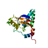
Mass: 18.015 Da / Num. of mol.: 26 / Source method: isolated from a natural source / Formula: H2O

-
Experimental details

-
Experiment

ExperimentMethod: X-RAY DIFFRACTION / Number of used crystals: 1

-
Sample preparation

CrystalDensity Matthews: 2.93 Å3/Da / Density % sol: 58.03 %
Crystal growTemperature: 293 K / Method: vapor diffusion, sitting drop / pH: 8.5 / Details: 0.1 M Tris-HCL, 0.7 M Na Citrate, pH 8.5

-
Data collection

DiffractionMean temperature: 100 K / Serial crystal experiment: N
Diffraction sourceSource: SYNCHROTRON / Site: APS / Beamline: 22-ID / Wavelength: 1 Å
DetectorType: DECTRIS EIGER X 16M / Detector: PIXEL / Date: Nov 19, 2019
RadiationProtocol: SINGLE WAVELENGTH / Monochromatic (M) / Laue (L): M / Scattering type: x-ray
Radiation wavelengthWavelength: 1 Å / Relative weight: 1
ReflectionResolution: 2.31→35.09 Å / Num. obs: 8574 / % possible obs: 98.8 % / Redundancy: 5.8 % / Biso Wilson estimate: 47.12 Å2 / CC1/2: 0.994 / Rmerge(I) obs: 0.171 / Rpim(I) all: 0.079 / Rrim(I) all: 0.189 / Net I/σ(I): 21
Reflection shellResolution: 2.31→2.45 Å / Redundancy: 5 % / Rmerge(I) obs: 0.832 / Mean I/σ(I) obs: 1.7 / Num. unique obs: 414 / CC1/2: 0.679 / Rpim(I) all: 0.391 / % possible all: 97.4

-
Processing

Software
NameVersionClassification
PHENIX1.14_3260refinement
HKL-2000data reduction
HKL-2000data scaling
PHASERphasing
RefinementMethod to determine structure: MOLECULAR REPLACEMENT
Starting model: 5XGH
Resolution: 2.31→35.09 Å / SU ML: 0.3327 / Cross valid method: FREE R-VALUE / σ(F): 1.34 / Phase error: 29.2057
RfactorNum. reflection% reflection
Rfree0.2552 855 10 %
Rwork0.1968 --
obs0.2027 8554 98.04 %
Solvent computationShrinkage radii: 0.9 Å / VDW probe radii: 1.11 Å
Displacement parametersBiso mean: 50.74 Å2
Refinement stepCycle: LAST / Resolution: 2.31→35.09 Å
ProteinNucleic acidLigandSolventTotal
Num. atoms1101 0 0 26 1127
Refine LS restraints
Refine-IDTypeDev idealNumber
X-RAY DIFFRACTIONf_bond_d0.01071134
X-RAY DIFFRACTIONf_angle_d0.95041538
X-RAY DIFFRACTIONf_chiral_restr0.0673174
X-RAY DIFFRACTIONf_plane_restr0.0059192
X-RAY DIFFRACTIONf_dihedral_angle_d19.1821430
LS refinement shell
Resolution (Å)Rfactor RfreeNum. reflection RfreeRfactor RworkNum. reflection RworkRefine-ID% reflection obs (%)
2.31-2.460.34841320.25651188X-RAY DIFFRACTION92.89
2.46-2.650.30581430.26281289X-RAY DIFFRACTION99.44
2.65-2.910.31791430.22881299X-RAY DIFFRACTION99.93
2.91-3.330.30491430.21531276X-RAY DIFFRACTION99.79
3.33-4.20.23211440.18621300X-RAY DIFFRACTION98.3
4.2-35.090.21621500.16971347X-RAY DIFFRACTION97.91

+
About Yorodumi

-
News

-
Feb 9, 2022. New format data for meta-information of EMDB entries

New format data for meta-information of EMDB entries

  • Version 3 of the EMDB header file is now the official format.
  • The previous official version 1.9 will be removed from the archive.

Related info.:EMDB header

External links:wwPDB to switch to version 3 of the EMDB data model

-
Aug 12, 2020. Covid-19 info

Covid-19 info

URL: https://pdbjlvh1.pdbj.org/emnavi/covid19.php

New page: Covid-19 featured information page in EM Navigator.

Related info.:Covid-19 info / Mar 5, 2020. Novel coronavirus structure data

+
Mar 5, 2020. Novel coronavirus structure data

Novel coronavirus structure data

Related info.:Yorodumi Speices / Aug 12, 2020. Covid-19 info

External links:COVID-19 featured content - PDBj / Molecule of the Month (242):Coronavirus Proteases

+
Jan 31, 2019. EMDB accession codes are about to change! (news from PDBe EMDB page)

EMDB accession codes are about to change! (news from PDBe EMDB page)

  • The allocation of 4 digits for EMDB accession codes will soon come to an end. Whilst these codes will remain in use, new EMDB accession codes will include an additional digit and will expand incrementally as the available range of codes is exhausted. The current 4-digit format prefixed with “EMD-” (i.e. EMD-XXXX) will advance to a 5-digit format (i.e. EMD-XXXXX), and so on. It is currently estimated that the 4-digit codes will be depleted around Spring 2019, at which point the 5-digit format will come into force.
  • The EM Navigator/Yorodumi systems omit the EMD- prefix.

Related info.:Q: What is EMD? / ID/Accession-code notation in Yorodumi/EM Navigator

External links:EMDB Accession Codes are Changing Soon! / Contact to PDBj

+
Jul 12, 2017. Major update of PDB

Major update of PDB

  • wwPDB released updated PDB data conforming to the new PDBx/mmCIF dictionary.
  • This is a major update changing the version number from 4 to 5, and with Remediation, in which all the entries are updated.
  • In this update, many items about electron microscopy experimental information are reorganized (e.g. em_software).
  • Now, EM Navigator and Yorodumi are based on the updated data.

External links:wwPDB Remediation / Enriched Model Files Conforming to OneDep Data Standards Now Available in the PDB FTP Archive

-
Yorodumi

Thousand views of thousand structures

  • Yorodumi is a browser for structure data from EMDB, PDB, SASBDB, etc.
  • This page is also the successor to EM Navigator detail page, and also detail information page/front-end page for Omokage search.
  • The word "yorodu" (or yorozu) is an old Japanese word meaning "ten thousand". "mi" (miru) is to see.

Related info.:EMDB / PDB / SASBDB / Comparison of 3 databanks / Yorodumi Search / Aug 31, 2016. New EM Navigator & Yorodumi / Yorodumi Papers / Jmol/JSmol / Function and homology information / Changes in new EM Navigator and Yorodumi

Read more