[English] 日本語
Yorodumi
- PDB-6nbu: CRISPR Complex Subunit Csm2 from Staphylococcus epidermidis RP62a -

+
Open data


ID or keywords:

Loading...

-
Basic information

Entry
Database: PDB / ID: 6nbu
TitleCRISPR Complex Subunit Csm2 from Staphylococcus epidermidis RP62a
ComponentsCRISPR-associated protein
KeywordsSTRUCTURAL PROTEIN / CRISPR
Function / homologyCRISPR-associated protein, Csm2 Type III-A / Csm2 Type III-A / defense response to virus / RNA binding / CRISPR system Cms protein Csm2
Function and homology information
Biological speciesStaphylococcus epidermidis (bacteria)
MethodX-RAY DIFFRACTION / SYNCHROTRON / SAD / Resolution: 2.75 Å
AuthorsDorsey, B.W. / Huang, L. / Mondragon, A.
Funding support United States, 1items
OrganizationGrant numberCountry
National Institutes of Health/National Institute of General Medical Sciences (NIH/NIGMS)R35 GM118108 United States
Citation
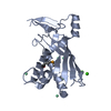
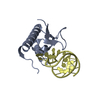
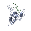
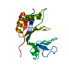
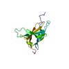
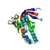
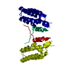
Journal: Nucleic Acids Res / Year: 2019
Title: Structural organization of a Type III-A CRISPR effector subcomplex determined by X-ray crystallography and cryo-EM.
Authors: Bryan W Dorsey / Lei Huang / Alfonso Mondragón /
Abstract: Clustered regularly interspaced short palindromic repeats (CRISPR) and their associated Cas proteins provide an immune-like response in many prokaryotes against extraneous nucleic acids. CRISPR-Cas ...Clustered regularly interspaced short palindromic repeats (CRISPR) and their associated Cas proteins provide an immune-like response in many prokaryotes against extraneous nucleic acids. CRISPR-Cas systems are classified into different classes and types. Class 1 CRISPR-Cas systems form multi-protein effector complexes that includes a guide RNA (crRNA) used to identify the target for destruction. Here we present crystal structures of Staphylococcus epidermidis Type III-A CRISPR subunits Csm2 and Csm3 and a 5.2 Å resolution single-particle cryo-electron microscopy (cryo-EM) reconstruction of an in vivo assembled effector subcomplex including the crRNA. The structures help to clarify the quaternary architecture of Type III-A effector complexes, and provide details on crRNA binding, target RNA binding and cleavage, and intermolecular interactions essential for effector complex assembly. The structures allow a better understanding of the organization of Type III-A CRISPR effector complexes as well as highlighting the overall similarities and differences with other Class 1 effector complexes.
History
DepositionDec 10, 2018Deposition site: RCSB / Processing site: RCSB
Revision 1.0Feb 13, 2019Provider: repository / Type: Initial release
Revision 1.1Feb 27, 2019Group: Data collection / Database references / Category: citation / citation_author
Item: _citation.country / _citation.journal_abbrev ..._citation.country / _citation.journal_abbrev / _citation.journal_id_ASTM / _citation.journal_id_CSD / _citation.journal_id_ISSN / _citation.pdbx_database_id_DOI / _citation.pdbx_database_id_PubMed / _citation.title / _citation.year / _citation_author.identifier_ORCID
Revision 1.2May 1, 2019Group: Data collection / Database references / Category: citation
Item: _citation.journal_volume / _citation.page_first / _citation.page_last
Revision 1.3Jan 1, 2020Group: Author supporting evidence / Category: pdbx_audit_support / Item: _pdbx_audit_support.funding_organization
Revision 1.4Mar 13, 2024Group: Data collection / Database references / Category: chem_comp_atom / chem_comp_bond / database_2
Item: _database_2.pdbx_DOI / _database_2.pdbx_database_accession

-
Structure visualization

Structure viewerMolecule:
MolmilJmol/JSmol

Downloads & links

-
Assembly

Deposited unit
A: CRISPR-associated protein


Theoretical massNumber of molelcules
Total (without water)18,1811
Polymers18,1811
Non-polymers00
Water0
1


  • Idetical with deposited unit
  • defined by author
TypeNameSymmetry operationNumber
identity operation1_555x,y,z1
Buried area0 Å2
ΔGint0 kcal/mol
Surface area8170 Å2
2
A: CRISPR-associated protein

A: CRISPR-associated protein


Theoretical massNumber of molelcules
Total (without water)36,3612
Polymers36,3612
Non-polymers00
Water0
TypeNameSymmetry operationNumber
identity operation1_555x,y,z1
crystal symmetry operation7_555y,x,-z1
Buried area2000 Å2
ΔGint-12 kcal/mol
Surface area14340 Å2
MethodPISA
Unit cell
Length a, b, c (Å)77.777, 77.777, 69.472
Angle α, β, γ (deg.)90.00, 90.00, 90.00
Int Tables number92
Space group name H-MP41212

-
Components

#1: Protein CRISPR-associated protein


Mass: 18180.727 Da / Num. of mol.: 1 / Fragment: Subunit Csm2
Source method: isolated from a genetically manipulated source
Source: (gene. exp.) Staphylococcus epidermidis (strain ATCC 35984 / RP62A) (bacteria)
Strain: ATCC 35984 / RP62A / Gene: SERP2460 / Plasmid: pMCSG7 / Production host: Escherichia coli (E. coli) / Strain (production host): Rosetta (DE3) / References: UniProt: Q5HK90

-
Experimental details

-
Experiment

ExperimentMethod: X-RAY DIFFRACTION / Number of used crystals: 1

-
Sample preparation

CrystalDensity Matthews: 2.89 Å3/Da / Density % sol: 57.43 %
Crystal growTemperature: 283 K / Method: vapor diffusion, hanging drop / pH: 7 / Details: Tris, ethanol

-
Data collection

DiffractionMean temperature: 100 K / Serial crystal experiment: N
Diffraction sourceSource: SYNCHROTRON / Site: APS / Beamline: 21-ID-G / Wavelength: 0.97856 Å
DetectorType: RAYONIX MX-300 / Detector: CCD / Date: Jun 9, 2011
RadiationMonochromator: C(111) / Protocol: SINGLE WAVELENGTH / Monochromatic (M) / Laue (L): M / Scattering type: x-ray
Radiation wavelengthWavelength: 0.97856 Å / Relative weight: 1
ReflectionResolution: 2.75→77.78 Å / Num. obs: 5931 / % possible obs: 99.9 % / Redundancy: 6.6 % / CC1/2: 0.999 / Net I/σ(I): 22.7
Reflection shellResolution: 2.75→2.88 Å / Redundancy: 5 % / Mean I/σ(I) obs: 1.8 / Num. unique obs: 765 / CC1/2: 0.645 / % possible all: 99.8

-
Processing

Software
NameVersionClassification
REFMAC5.8.0232refinement
XDSdata reduction
Aimlessdata scaling
SHARPphasing
RefinementMethod to determine structure: SAD / Resolution: 2.75→25 Å / Cor.coef. Fo:Fc: 0.939 / Cor.coef. Fo:Fc free: 0.921 / Cross valid method: THROUGHOUT / ESU R: 0.609 / ESU R Free: 0.358 / Details: HYDROGENS HAVE BEEN ADDED IN THE RIDING POSITIONS
RfactorNum. reflection% reflectionSelection details
Rfree0.29418 256 4.3 %RANDOM
Rwork0.24471 ---
obs0.24689 5635 99.71 %-
Solvent computationIon probe radii: 0.8 Å / Shrinkage radii: 0.8 Å / VDW probe radii: 1.2 Å
Displacement parametersBiso mean: 76.195 Å2
Baniso -1Baniso -2Baniso -3
1--0.59 Å20 Å20 Å2
2---0.59 Å20 Å2
3---1.19 Å2
Refinement stepCycle: 1 / Resolution: 2.75→25 Å
ProteinNucleic acidLigandSolventTotal
Num. atoms1071 0 0 0 1071
Refine LS restraints
Refine-IDTypeDev idealDev ideal targetNumber
X-RAY DIFFRACTIONr_bond_refined_d0.0140.0131092
X-RAY DIFFRACTIONr_bond_other_d0.0340.0171018
X-RAY DIFFRACTIONr_angle_refined_deg1.6891.6481463
X-RAY DIFFRACTIONr_angle_other_deg2.2221.5822366
X-RAY DIFFRACTIONr_dihedral_angle_1_deg5.5095124
X-RAY DIFFRACTIONr_dihedral_angle_2_deg37.33523.96863
X-RAY DIFFRACTIONr_dihedral_angle_3_deg17.10115212
X-RAY DIFFRACTIONr_dihedral_angle_4_deg9.987154
X-RAY DIFFRACTIONr_chiral_restr0.1160.2135
X-RAY DIFFRACTIONr_gen_planes_refined0.0040.021188
X-RAY DIFFRACTIONr_gen_planes_other0.0050.02240
X-RAY DIFFRACTIONr_nbd_refined
X-RAY DIFFRACTIONr_nbd_other
X-RAY DIFFRACTIONr_nbtor_refined
X-RAY DIFFRACTIONr_nbtor_other
X-RAY DIFFRACTIONr_xyhbond_nbd_refined
X-RAY DIFFRACTIONr_xyhbond_nbd_other
X-RAY DIFFRACTIONr_metal_ion_refined
X-RAY DIFFRACTIONr_metal_ion_other
X-RAY DIFFRACTIONr_symmetry_vdw_refined
X-RAY DIFFRACTIONr_symmetry_vdw_other
X-RAY DIFFRACTIONr_symmetry_hbond_refined
X-RAY DIFFRACTIONr_symmetry_hbond_other
X-RAY DIFFRACTIONr_symmetry_metal_ion_refined
X-RAY DIFFRACTIONr_symmetry_metal_ion_other
X-RAY DIFFRACTIONr_mcbond_it4.0298.118502
X-RAY DIFFRACTIONr_mcbond_other4.0088.112501
X-RAY DIFFRACTIONr_mcangle_it5.60712.151624
X-RAY DIFFRACTIONr_mcangle_other5.60212.158625
X-RAY DIFFRACTIONr_scbond_it5.0938.459590
X-RAY DIFFRACTIONr_scbond_other5.0898.46591
X-RAY DIFFRACTIONr_scangle_it
X-RAY DIFFRACTIONr_scangle_other6.71812.547840
X-RAY DIFFRACTIONr_long_range_B_refined7.33393.351297
X-RAY DIFFRACTIONr_long_range_B_other7.33893.4121298
X-RAY DIFFRACTIONr_rigid_bond_restr
X-RAY DIFFRACTIONr_sphericity_free
X-RAY DIFFRACTIONr_sphericity_bonded
LS refinement shellResolution: 2.75→2.821 Å / Total num. of bins used: 20
RfactorNum. reflection% reflection
Rfree0.367 22 -
Rwork0.323 387 -
obs--99.03 %

+
About Yorodumi

-
News

-
Feb 9, 2022. New format data for meta-information of EMDB entries

New format data for meta-information of EMDB entries

  • Version 3 of the EMDB header file is now the official format.
  • The previous official version 1.9 will be removed from the archive.

Related info.:EMDB header

External links:wwPDB to switch to version 3 of the EMDB data model

-
Aug 12, 2020. Covid-19 info

Covid-19 info

URL: https://pdbj.org/emnavi/covid19.php

New page: Covid-19 featured information page in EM Navigator.

Related info.:Covid-19 info / Mar 5, 2020. Novel coronavirus structure data

+
Mar 5, 2020. Novel coronavirus structure data

Novel coronavirus structure data

Related info.:Yorodumi Speices / Aug 12, 2020. Covid-19 info

External links:COVID-19 featured content - PDBj / Molecule of the Month (242):Coronavirus Proteases

+
Jan 31, 2019. EMDB accession codes are about to change! (news from PDBe EMDB page)

EMDB accession codes are about to change! (news from PDBe EMDB page)

  • The allocation of 4 digits for EMDB accession codes will soon come to an end. Whilst these codes will remain in use, new EMDB accession codes will include an additional digit and will expand incrementally as the available range of codes is exhausted. The current 4-digit format prefixed with “EMD-” (i.e. EMD-XXXX) will advance to a 5-digit format (i.e. EMD-XXXXX), and so on. It is currently estimated that the 4-digit codes will be depleted around Spring 2019, at which point the 5-digit format will come into force.
  • The EM Navigator/Yorodumi systems omit the EMD- prefix.

Related info.:Q: What is EMD? / ID/Accession-code notation in Yorodumi/EM Navigator

External links:EMDB Accession Codes are Changing Soon! / Contact to PDBj

+
Jul 12, 2017. Major update of PDB

Major update of PDB

  • wwPDB released updated PDB data conforming to the new PDBx/mmCIF dictionary.
  • This is a major update changing the version number from 4 to 5, and with Remediation, in which all the entries are updated.
  • In this update, many items about electron microscopy experimental information are reorganized (e.g. em_software).
  • Now, EM Navigator and Yorodumi are based on the updated data.

External links:wwPDB Remediation / Enriched Model Files Conforming to OneDep Data Standards Now Available in the PDB FTP Archive

-
Yorodumi

Thousand views of thousand structures

  • Yorodumi is a browser for structure data from EMDB, PDB, SASBDB, etc.
  • This page is also the successor to EM Navigator detail page, and also detail information page/front-end page for Omokage search.
  • The word "yorodu" (or yorozu) is an old Japanese word meaning "ten thousand". "mi" (miru) is to see.

Related info.:EMDB / PDB / SASBDB / Comparison of 3 databanks / Yorodumi Search / Aug 31, 2016. New EM Navigator & Yorodumi / Yorodumi Papers / Jmol/JSmol / Function and homology information / Changes in new EM Navigator and Yorodumi

Read more