| Entry | Database: PDB / ID: 5xgh
|
|---|
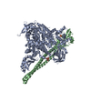
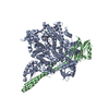
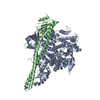
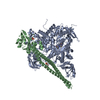
| Title | Crystal structure of PI3K complex with an inhibitor |
|---|
 Components Components | (Phosphatidylinositol ...) x 2 |
|---|
 Keywords Keywords | TRANSFERASE/INHIBITOR / PI3K / inhibitor / TRANSFERASE-INHIBITOR complex |
|---|
| Function / homology |  Function and homology information Function and homology information
perinuclear endoplasmic reticulum membrane / regulation of toll-like receptor 4 signaling pathway / response to muscle inactivity / phosphatidylinositol kinase activity / phosphatidylinositol 3-kinase regulator activity / 1-phosphatidylinositol-3-kinase regulator activity / positive regulation of endoplasmic reticulum unfolded protein response / regulation of actin filament organization / negative regulation of actin filament depolymerization / phosphatidylinositol 3-kinase activator activity ...perinuclear endoplasmic reticulum membrane / regulation of toll-like receptor 4 signaling pathway / response to muscle inactivity / phosphatidylinositol kinase activity / phosphatidylinositol 3-kinase regulator activity / 1-phosphatidylinositol-3-kinase regulator activity / positive regulation of endoplasmic reticulum unfolded protein response / regulation of actin filament organization / negative regulation of actin filament depolymerization / phosphatidylinositol 3-kinase activator activity / response to butyrate / T follicular helper cell differentiation / IRS-mediated signalling / interleukin-18-mediated signaling pathway / phosphatidylinositol 3-kinase regulatory subunit binding / response to L-leucine / PI3K events in ERBB4 signaling / neurotrophin TRKA receptor binding / positive regulation of focal adhesion disassembly / cellular response to hydrostatic pressure / cis-Golgi network / Dengue Virus Attachment and Entry / Activated NTRK2 signals through PI3K / ErbB-3 class receptor binding / negative regulation of stress fiber assembly / transmembrane receptor protein tyrosine kinase adaptor activity / Activated NTRK3 signals through PI3K / phosphatidylinositol 3-kinase complex, class IB / phosphatidylinositol 3-kinase complex / TORC2 signaling / Co-stimulation by ICOS / RHOD GTPase cycle / positive regulation of protein localization to membrane / Signaling by cytosolic FGFR1 fusion mutants / vasculature development / Nephrin family interactions / RHOF GTPase cycle / kinase activator activity / Signaling by LTK in cancer / 1-phosphatidylinositol-4-phosphate 3-kinase activity / Signaling by LTK / anoikis / RND1 GTPase cycle / phosphatidylinositol 3-kinase complex, class IA / positive regulation of filopodium assembly / RND2 GTPase cycle / MET activates PI3K/AKT signaling / PI3K/AKT activation / relaxation of cardiac muscle / RND3 GTPase cycle / phosphatidylinositol-4,5-bisphosphate 3-kinase / 1-phosphatidylinositol-4,5-bisphosphate 3-kinase activity / growth hormone receptor signaling pathway / insulin binding / phosphatidylinositol 3-kinase / natural killer cell mediated cytotoxicity / phosphatidylinositol-3-phosphate biosynthetic process / Signaling by ALK / cardiac muscle cell contraction / RHOV GTPase cycle / 1-phosphatidylinositol-3-kinase activity / vascular endothelial growth factor signaling pathway / RHOB GTPase cycle / negative regulation of macroautophagy / GP1b-IX-V activation signalling / Erythropoietin activates Phosphoinositide-3-kinase (PI3K) / PI-3K cascade:FGFR3 / response to dexamethasone / PI-3K cascade:FGFR2 / PI-3K cascade:FGFR4 / positive regulation of protein kinase activity / PI-3K cascade:FGFR1 / RHOJ GTPase cycle / RHOC GTPase cycle / phosphatidylinositol phosphate biosynthetic process / Synthesis of PIPs at the plasma membrane / RHOU GTPase cycle / phosphatidylinositol-mediated signaling / CDC42 GTPase cycle / RET signaling / negative regulation of anoikis / Interleukin-3, Interleukin-5 and GM-CSF signaling / T cell differentiation / PI3K events in ERBB2 signaling / RHOG GTPase cycle / insulin receptor substrate binding / intercalated disc / PI3K Cascade / Role of LAT2/NTAL/LAB on calcium mobilization / RAC3 GTPase cycle / RHOA GTPase cycle / CD28 dependent PI3K/Akt signaling / RAC2 GTPase cycle / Interleukin receptor SHC signaling / positive regulation of TOR signaling / Role of phospholipids in phagocytosis / enzyme-substrate adaptor activity / GAB1 signalosome / protein kinase activator activity / endothelial cell migrationSimilarity search - Function Helix Hairpins - #1490 / Phosphatidylinositol 3-kinase regulatory subunit alpha, SH3 domain / PIK3R1, inter-SH2 domain / PI3Kalpha, catalytic domain / PI3K p85 subunit, C-terminal SH2 domain / PI3K regulatory subunit p85-related , inter-SH2 domain / PI3K p85 subunit, N-terminal SH2 domain / Phosphatidylinositol 3-kinase regulatory subunit P85 inter-SH2 domain / PI3-kinase family, p85-binding domain / PI3-kinase family, p85-binding domain ...Helix Hairpins - #1490 / Phosphatidylinositol 3-kinase regulatory subunit alpha, SH3 domain / PIK3R1, inter-SH2 domain / PI3Kalpha, catalytic domain / PI3K p85 subunit, C-terminal SH2 domain / PI3K regulatory subunit p85-related , inter-SH2 domain / PI3K p85 subunit, N-terminal SH2 domain / Phosphatidylinositol 3-kinase regulatory subunit P85 inter-SH2 domain / PI3-kinase family, p85-binding domain / PI3-kinase family, p85-binding domain / Phosphatidylinositol 3-kinase, accessory domain (PIK) / Rho GTPase-activating protein domain / RhoGAP domain / Rho GTPase-activating proteins domain profile. / GTPase-activator protein for Rho-like GTPases / Phosphatidylinositol 3-kinase, adaptor-binding domain / Phosphatidylinositol 3-kinase adaptor-binding (PI3K ABD) domain profile. / PI3-kinase family, Ras-binding domain / Phosphatidylinositol 3-kinase Ras-binding (PI3K RBD) domain / PI3-kinase family, ras-binding domain / Phosphatidylinositol 3-kinase Ras-binding (PI3K RBD) domain profile. / Rho GTPase activation protein / C2 domain / SH2 domain / C2 phosphatidylinositol 3-kinase-type domain / Phosphoinositide 3-kinase C2 / C2 phosphatidylinositol 3-kinase (PI3K)-type domain profile. / Phosphoinositide 3-kinase, region postulated to contain C2 domain / SHC Adaptor Protein / Phosphoinositide 3-kinase family, accessory domain (PIK domain) / Phosphoinositide 3-kinase family, accessory domain (PIK domain) / Phosphoinositide 3-kinase, accessory (PIK) domain superfamily / Phosphoinositide 3-kinase, accessory (PIK) domain / Phosphatidylinositol kinase / PIK helical domain profile. / Phosphatidylinositol 3- and 4-kinases signature 1. / Phosphatidylinositol 3/4-kinase, conserved site / Phosphatidylinositol 3- and 4-kinases signature 2. / Phosphatidylinositol 3-/4-kinase, catalytic domain superfamily / Phosphoinositide 3-kinase, catalytic domain / Phosphatidylinositol 3- and 4-kinase / Phosphatidylinositol 3- and 4-kinases catalytic domain profile. / Phosphatidylinositol 3-/4-kinase, catalytic domain / C2 domain superfamily / SH2 domain / Src homology 2 (SH2) domain profile. / Src homology 2 domains / SH2 domain / Src homology 3 domains / SH2 domain superfamily / Serine Threonine Protein Phosphatase 5, Tetratricopeptide repeat / SH3-like domain superfamily / Src homology 3 (SH3) domain profile. / SH3 domain / Helix Hairpins / Alpha Horseshoe / Armadillo-type fold / Ubiquitin-like domain superfamily / Protein kinase-like domain superfamily / Immunoglobulin-like / Sandwich / 2-Layer Sandwich / Orthogonal Bundle / Mainly Beta / Mainly Alpha / Alpha BetaSimilarity search - Domain/homology Chem-84U / Phosphatidylinositol 3-kinase regulatory subunit alpha / Phosphatidylinositol 4,5-bisphosphate 3-kinase catalytic subunit alpha isoformSimilarity search - Component |
|---|
| Biological species |  Homo sapiens (human) Homo sapiens (human) |
|---|
| Method |  X-RAY DIFFRACTION / SYNCHROTRON / MOLECULAR REPLACEMENT / Resolution: 2.97 Å X-RAY DIFFRACTION / SYNCHROTRON / MOLECULAR REPLACEMENT / Resolution: 2.97 Å |
|---|
 Authors Authors | Song, K. / Yang, X. / Zhao, Y. / Jian, Z. |
|---|
 Citation Citation | Journal: Sci Rep / Year: 2017
Title: New Insights into PI3K Inhibitor Design using X-ray Structures of PI3K alpha Complexed with a Potent Lead Compound.
Authors: Yang, X. / Zhang, X. / Huang, M. / Song, K. / Li, X. / Huang, M. / Meng, L. / Zhang, J. |
|---|
| History | | Deposition | Apr 13, 2017 | Deposition site: PDBJ / Processing site: PDBJ |
|---|
| Revision 1.0 | Apr 25, 2018 | Provider: repository / Type: Initial release |
|---|
| Revision 1.1 | Nov 6, 2019 | Group: Data collection / Database references / Category: citation / citation_author
Item: _citation.country / _citation.journal_abbrev ..._citation.country / _citation.journal_abbrev / _citation.journal_id_CSD / _citation.journal_id_ISSN / _citation.journal_volume / _citation.page_first / _citation.page_last / _citation.pdbx_database_id_DOI / _citation.pdbx_database_id_PubMed / _citation.title / _citation.year |
|---|
| Revision 1.2 | Mar 27, 2024 | Group: Data collection / Database references / Category: chem_comp_atom / chem_comp_bond / database_2
Item: _database_2.pdbx_DOI / _database_2.pdbx_database_accession |
|---|
|
|---|
 Movie
Movie Controller


 Open data
 Basic information
 Structure visualization
 Downloads & links
 Other downloads


 PDBj
PDBj


 Assembly

 Spodoptera frugiperda (fall armyworm)


 Mass: 436.502 Da / Num. of mol.: 1 / Source method: obtained synthetically / Formula: C23H21FN4O2S
 Mass: 92.094 Da / Num. of mol.: 1 / Source method: obtained synthetically / Formula: C3H8O3
 Mass: 96.063 Da / Num. of mol.: 1 / Source method: obtained synthetically / Formula: SO4
 Sample preparation
 / Beamline: BL19U1 / Wavelength: 1 Å
 Processing