 Movie
Movie Controller


+ Open data

- Basic information
| Entry | Database: PDB / ID: 6vjg | |||||||||
|---|---|---|---|---|---|---|---|---|---|---|
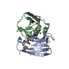
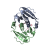
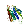
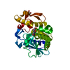
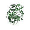
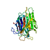
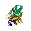
| Title | Csx3-I222 Crystal Form at 1.8 Angstrom Resolution | |||||||||
 Components Components | CRISPR-associated protein, Csx3 family | |||||||||
 Keywords Keywords | RNA BINDING PROTEIN / CRISPR / Cas / CARF / RNase | |||||||||
| Function / homology | CRISPR-associated protein Csx3 / CRISPR-associated protein (Cas_csx3) / CRISPR-associated protein, Csx3 family / Uncharacterized protein AF_1864 Function and homology information Function and homology information | |||||||||
| Biological species |   Archaeoglobus fulgidus DSM 8774 (archaea) Archaeoglobus fulgidus DSM 8774 (archaea) | |||||||||
| Method |  X-RAY DIFFRACTION / SYNCHROTRON / MOLECULAR REPLACEMENT / Resolution: 1.8 Å X-RAY DIFFRACTION / SYNCHROTRON / MOLECULAR REPLACEMENT / Resolution: 1.8 Å | |||||||||
 Authors Authors | Brown, S. / Charbonneau, A. / Burman, N. / Gauvin, C.C. / Lawrence, C.M. | |||||||||
| Funding support |  United States, 2items United States, 2items
| |||||||||
 Citation Citation | Journal: J.Biol.Chem. / Year: 2020 Title: Csx3 is a cyclic oligonucleotide phosphodiesterase associated with type III CRISPR-Cas that degrades the second messenger cA 4 . Authors: Brown, S. / Gauvin, C.C. / Charbonneau, A.A. / Burman, N. / Lawrence, C.M. | |||||||||
| History |
|
- Structure visualization
| Structure viewer | Molecule: MolmilJmol/JSmol |
|---|
- Downloads & links
-Download
| PDBx/mmCIF format | 6vjg.cif.gz | 69.6 KB | Display | PDBx/mmCIF format |
|---|---|---|---|---|
| PDB format | pdb6vjg.ent.gz | 42 KB | Display | PDB format |
| PDBx/mmJSON format | 6vjg.json.gz | Tree view | PDBx/mmJSON format | |
| Others |  Other downloads Other downloads |
-Validation report
| Arichive directory | https://data.pdbj.org/pub/pdb/validation_reports/vj/6vjgftp://data.pdbj.org/pub/pdb/validation_reports/vj/6vjg | HTTPS FTP |
|---|
-Related structure data
| Related structure data | 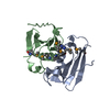 3wzgS S: Starting model for refinement |
|---|---|
| Similar structure data |


-Links
 PDBj
PDBj
- Assembly
| Deposited unit | 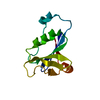
| ||||||||||||
|---|---|---|---|---|---|---|---|---|---|---|---|---|---|
| 1 | 
| ||||||||||||
| Unit cell |
| ||||||||||||
| Components on special symmetry positions |
|
-Components
| #1: Protein | Mass: 12694.774 Da / Num. of mol.: 1 Source method: isolated from a genetically manipulated source Source: (gene. exp.) Archaeoglobus fulgidus DSM 8774 (archaea)Gene: AFULGI_00021190 / Production host:  |
|---|---|
| #2: Water | ChemComp-HOH /  Mass: 18.015 Da / Num. of mol.: 54 / Source method: isolated from a natural source / Formula: H2O Mass: 18.015 Da / Num. of mol.: 54 / Source method: isolated from a natural source / Formula: H2O |
-Experimental details
-Experiment
| Experiment | Method: X-RAY DIFFRACTION / Number of used crystals: 1 |
|---|
- Sample preparation
| Crystal | Density Matthews: 2.43 Å3/Da / Density % sol: 45.94 % Description: Rhombus shaped plates approximately 115 x 125 um. |
|---|---|
| Crystal grow | Temperature: 295 K / Method: vapor diffusion, hanging drop / pH: 8.5 Details: 45% 2-methyl-2,4-pentanediol, 0.1 M Tris pH 8.5, 0.2 M Ammonium Phosphate Monobasic |
-Data collection
| Diffraction | Mean temperature: 100 K / Serial crystal experiment: N |
|---|---|
| Diffraction source | Source: SYNCHROTRON / Site: APS / Beamline: 24-ID-C / Wavelength: 0.979 Å |
| Detector | Type: DECTRIS PILATUS 6M-F / Detector: PIXEL / Date: Oct 23, 2019 |
| Radiation | Protocol: SINGLE WAVELENGTH / Monochromatic (M) / Laue (L): M / Scattering type: x-ray |
| Radiation wavelength | Wavelength: 0.979 Å / Relative weight: 1 |
| Reflection | Resolution: 1.8→46.29 Å / Num. obs: 10996 / % possible obs: 98.9 % / Redundancy: 4.8 % / CC1/2: 0.999 / CC star: 1 / Rmerge(I) obs: 0.034 / Rpim(I) all: 0.032 / Rrim(I) all: 0.071 / Χ2: 0.933 / Net I/σ(I): 27.305 |
| Reflection shell | Resolution: 1.8→1.83 Å / Redundancy: 4.1 % / Rmerge(I) obs: 0.439 / Mean I/σ(I) obs: 2.87 / Num. unique obs: 510 / CC1/2: 0.917 / CC star: 0.978 / Χ2: 0.53 / % possible all: 96.4 |
- Processing
| Software |
| |||||||||||||||||||||||||||||||||||||||||||||||||||||||||||||||
|---|---|---|---|---|---|---|---|---|---|---|---|---|---|---|---|---|---|---|---|---|---|---|---|---|---|---|---|---|---|---|---|---|---|---|---|---|---|---|---|---|---|---|---|---|---|---|---|---|---|---|---|---|---|---|---|---|---|---|---|---|---|---|---|---|
| Refinement | Method to determine structure: MOLECULAR REPLACEMENT Starting model: 3WZG Resolution: 1.8→46.25 Å / SU ML: 0.1904 / Cross valid method: FREE R-VALUE / σ(F): 1.38 / Phase error: 25.0208
| |||||||||||||||||||||||||||||||||||||||||||||||||||||||||||||||
| Solvent computation | Shrinkage radii: 0.9 Å / VDW probe radii: 1.11 Å | |||||||||||||||||||||||||||||||||||||||||||||||||||||||||||||||
| Displacement parameters | Biso mean: 45.68 Å2 | |||||||||||||||||||||||||||||||||||||||||||||||||||||||||||||||
| Refinement step | Cycle: LAST / Resolution: 1.8→46.25 Å
| |||||||||||||||||||||||||||||||||||||||||||||||||||||||||||||||
| Refine LS restraints |
| |||||||||||||||||||||||||||||||||||||||||||||||||||||||||||||||
| LS refinement shell |
| |||||||||||||||||||||||||||||||||||||||||||||||||||||||||||||||
| Refinement TLS params. | Method: refined / Origin x: 3.95343482676 Å / Origin y: 14.817631476 Å / Origin z: 9.75784545109 Å
| |||||||||||||||||||||||||||||||||||||||||||||||||||||||||||||||
| Refinement TLS group | Selection details: all |
