 Movie
Movie Controller


+ Open data

- Basic information
| Entry | Database: PDB / ID: 6v6c | |||||||||||||||||||||||||||||||||
|---|---|---|---|---|---|---|---|---|---|---|---|---|---|---|---|---|---|---|---|---|---|---|---|---|---|---|---|---|---|---|---|---|---|---|
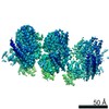
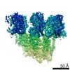
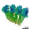
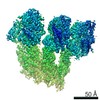
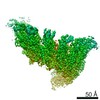
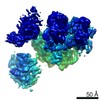
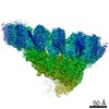
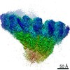
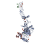
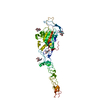
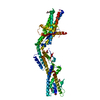
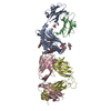
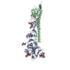
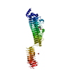
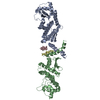
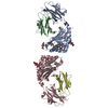
| Title | Structure of GCP6 in the native human gamma-tubulin ring complex | |||||||||||||||||||||||||||||||||
 Components Components | Gamma-tubulin complex component 6 | |||||||||||||||||||||||||||||||||
 Keywords Keywords | STRUCTURAL PROTEIN / GCP / GCP6 / gamma-tubulin ring complex / gTuRC / g-TuRC / microtubule / microtubule nucleation / single particle cryo-EM structure | |||||||||||||||||||||||||||||||||
| Function / homology |  Function and homology information Function and homology informationgamma-tubulin complex / gamma-tubulin ring complex / microtubule nucleation / gamma-tubulin binding / spindle assembly / cytoplasmic microtubule organization / Recruitment of mitotic centrosome proteins and complexes / Recruitment of NuMA to mitotic centrosomes / meiotic cell cycle / spindle pole ...gamma-tubulin complex / gamma-tubulin ring complex / microtubule nucleation / gamma-tubulin binding / spindle assembly / cytoplasmic microtubule organization / Recruitment of mitotic centrosome proteins and complexes / Recruitment of NuMA to mitotic centrosomes / meiotic cell cycle / spindle pole / mitotic cell cycle / microtubule binding / microtubule / centrosome / membrane / cytosol Similarity search - Function | |||||||||||||||||||||||||||||||||
| Biological species |  Homo sapiens (human) Homo sapiens (human) | |||||||||||||||||||||||||||||||||
| Method | ELECTRON MICROSCOPY / single particle reconstruction / cryo EM / Resolution: 4.5 Å | |||||||||||||||||||||||||||||||||
 Authors Authors | Wieczorek, M. / Urnavicius, L. / Ti, S. / Molloy, K.R. / Chait, B.T. / Kapoor, T.M. | |||||||||||||||||||||||||||||||||
| Funding support |  United States, United States,  France, 2items France, 2items
| |||||||||||||||||||||||||||||||||
 Citation Citation | Journal: Cell / Year: 2020 Title: Asymmetric Molecular Architecture of the Human γ-Tubulin Ring Complex. Authors: Michal Wieczorek / Linas Urnavicius / Shih-Chieh Ti / Kelly R Molloy / Brian T Chait / Tarun M Kapoor / Abstract: The γ-tubulin ring complex (γ-TuRC) is an essential regulator of centrosomal and acentrosomal microtubule formation, yet its structure is not known. Here, we present a cryo-EM reconstruction of the ...The γ-tubulin ring complex (γ-TuRC) is an essential regulator of centrosomal and acentrosomal microtubule formation, yet its structure is not known. Here, we present a cryo-EM reconstruction of the native human γ-TuRC at ∼3.8 Å resolution, revealing an asymmetric, cone-shaped structure. Pseudo-atomic models indicate that GCP4, GCP5, and GCP6 form distinct Y-shaped assemblies that structurally mimic GCP2/GCP3 subcomplexes distal to the γ-TuRC "seam." We also identify an unanticipated structural bridge that includes an actin-like protein and spans the γ-TuRC lumen. Despite its asymmetric architecture, the γ-TuRC arranges γ-tubulins into a helical geometry poised to nucleate microtubules. Diversity in the γ-TuRC subunits introduces large (>100,000 Å) surfaces in the complex that allow for interactions with different regulatory factors. The observed compositional complexity of the γ-TuRC could self-regulate its assembly into a cone-shaped structure to control microtubule formation across diverse contexts, e.g., within biological condensates or alongside existing filaments. | |||||||||||||||||||||||||||||||||
| History |
|
- Structure visualization
| Movie |
Movie viewer |
|---|---|
| Structure viewer | Molecule: MolmilJmol/JSmol |
- Downloads & links
-Download
| PDBx/mmCIF format | 6v6c.cif.gz | 129.6 KB | Display | PDBx/mmCIF format |
|---|---|---|---|---|
| PDB format | pdb6v6c.ent.gz | 81.5 KB | Display | PDB format |
| PDBx/mmJSON format | 6v6c.json.gz | Tree view | PDBx/mmJSON format | |
| Others |  Other downloads Other downloads |
-Validation report
| Arichive directory | https://data.pdbj.org/pub/pdb/validation_reports/v6/6v6cftp://data.pdbj.org/pub/pdb/validation_reports/v6/6v6c | HTTPS FTP |
|---|
-Related structure data
| Related structure data | 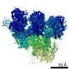 21068MC 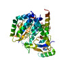 6v5vC 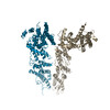 6v69C 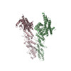 6v6bC 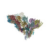 6v6sC M: map data used to model this data C: citing same article ( |
|---|---|
| Similar structure data |


-Links
 PDBj
PDBj
- Assembly
| Deposited unit | 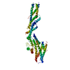
|
|---|---|
| 1 |
|
-Components
| #1: Protein | Mass: 200733.641 Da / Num. of mol.: 1 / Source method: isolated from a natural source Details: Please note that the full sequence for GCP6 (Uniprot accession number Q96RT7) was much longer than our modeled region, and the alignment failed to capture the domains we built, despite all ...Details: Please note that the full sequence for GCP6 (Uniprot accession number Q96RT7) was much longer than our modeled region, and the alignment failed to capture the domains we built, despite all residues being numbered accordingly in the submitted coordinates. See below for full sequence. Source: (natural) Homo sapiens (human) / References: UniProt: Q96RT7 |
|---|---|
| Has protein modification | N |
-Experimental details
-Experiment
| Experiment | Method: ELECTRON MICROSCOPY |
|---|---|
| EM experiment | Aggregation state: PARTICLE / 3D reconstruction method: single particle reconstruction |
- Sample preparation
| Component | Name: Native human gamma-tubulin ring complex / Type: COMPLEX / Entity ID: all / Source: NATURAL |
|---|---|
| Molecular weight | Experimental value: NO |
| Source (natural) | Organism: Homo sapiens (human) |
| Buffer solution | pH: 7.5 |
| Specimen | Embedding applied: NO / Shadowing applied: NO / Staining applied: NO / Vitrification applied: YES |
| Vitrification | Cryogen name: ETHANE |
- Electron microscopy imaging
| Experimental equipment |  Model: Titan Krios / Image courtesy: FEI Company |
|---|---|
| Microscopy | Model: FEI TITAN KRIOS |
| Electron gun | Electron source:  FIELD EMISSION GUN / Accelerating voltage: 300 kV / Illumination mode: OTHER FIELD EMISSION GUN / Accelerating voltage: 300 kV / Illumination mode: OTHER |
| Electron lens | Mode: BRIGHT FIELD |
| Image recording | Electron dose: 45 e/Å2 / Film or detector model: GATAN K2 SUMMIT (4k x 4k) |
- Processing
| Software | Name: PHENIX / Version: 1.16_3549: / Classification: refinement | ||||||||||||||||||||||||
|---|---|---|---|---|---|---|---|---|---|---|---|---|---|---|---|---|---|---|---|---|---|---|---|---|---|
| EM software | Name: PHENIX / Category: model refinement | ||||||||||||||||||||||||
| CTF correction | Type: PHASE FLIPPING AND AMPLITUDE CORRECTION | ||||||||||||||||||||||||
| Symmetry | Point symmetry: C1 (asymmetric) | ||||||||||||||||||||||||
| 3D reconstruction | Resolution: 4.5 Å / Resolution method: FSC 0.143 CUT-OFF / Num. of particles: 102613 / Symmetry type: POINT | ||||||||||||||||||||||||
| Refine LS restraints |
|
