 Movie
Movie Controller

[English] 日本語
 Yorodumi
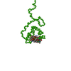
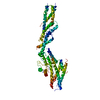
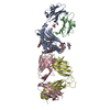
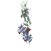
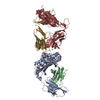
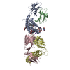
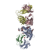
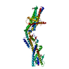
- PDB-6fkq: THE CRYSTAL STRUCTURE OF A FRAGMENT OF NETRIN-1 IN COMPLEX WITH A... -
+ Open data

- Basic information
| Entry | Database: PDB / ID: 6fkq | ||||||||||||
|---|---|---|---|---|---|---|---|---|---|---|---|---|---|
| Title | THE CRYSTAL STRUCTURE OF A FRAGMENT OF NETRIN-1 IN COMPLEX WITH A FRAGMENT OF DRAXIN | ||||||||||||
 Components Components |
| ||||||||||||
 Keywords Keywords | SIGNALING PROTEIN / AXON GUIDANCE CUE / NETRIN-1 / DRAXIN / COMMISSURAL NEURON | ||||||||||||
| Function / homology |  Function and homology information Function and homology informationcommissural neuron differentiation in spinal cord / dorsal spinal cord development / anterior commissure morphogenesis / regulation of glial cell migration / DSCAM interactions / chemorepulsion of axon / Cdc42 protein signal transduction / anterior/posterior axon guidance / negative regulation of hippocampal neuron apoptotic process / Netrin-1 signaling ...commissural neuron differentiation in spinal cord / dorsal spinal cord development / anterior commissure morphogenesis / regulation of glial cell migration / DSCAM interactions / chemorepulsion of axon / Cdc42 protein signal transduction / anterior/posterior axon guidance / negative regulation of hippocampal neuron apoptotic process / Netrin-1 signaling / motor neuron migration / Regulation of commissural axon pathfinding by SLIT and ROBO / Role of second messengers in netrin-1 signaling / negative regulation of axon extension / mammary gland duct morphogenesis / Netrin mediated repulsion signals / substrate-dependent cell migration, cell extension / DCC mediated attractive signaling / hippocampal neuron apoptotic process / positive regulation of cell motility / inner ear morphogenesis / nuclear migration / regulation of synapse assembly / forebrain development / basement membrane / positive regulation of glial cell proliferation / positive regulation of axon extension / glial cell proliferation / axon guidance / negative regulation of canonical Wnt signaling pathway / cell-cell adhesion / Wnt signaling pathway / actin cytoskeleton / molecular adaptor activity / DNA-binding transcription factor activity, RNA polymerase II-specific / Ras protein signal transduction / RNA polymerase II cis-regulatory region sequence-specific DNA binding / apoptotic process / regulation of transcription by RNA polymerase II / glutamatergic synapse / extracellular region / nucleoplasm / cytosol Similarity search - Function | ||||||||||||
| Biological species |  Homo sapiens (human) Homo sapiens (human) | ||||||||||||
| Method |  X-RAY DIFFRACTION / SYNCHROTRON / MOLECULAR REPLACEMENT / Resolution: 3.07 Å X-RAY DIFFRACTION / SYNCHROTRON / MOLECULAR REPLACEMENT / Resolution: 3.07 Å | ||||||||||||
 Authors Authors | Bhowmick, T. / Meijers, R. | ||||||||||||
| Funding support |  Germany, 1items Germany, 1items
| ||||||||||||
 Citation Citation | Journal: Neuron / Year: 2018 Title: Structural Basis for Draxin-Modulated Axon Guidance and Fasciculation by Netrin-1 through DCC. Authors: Ying Liu / Tuhin Bhowmick / Yiqiong Liu / Xuefan Gao / Haydyn D T Mertens / Dmitri I Svergun / Junyu Xiao / Yan Zhang / Jia-Huai Wang / Rob Meijers /   Abstract: Axon guidance involves the spatiotemporal interplay between guidance cues and membrane-bound cell-surface receptors, present on the growth cone of the axon. Netrin-1 is a prototypical guidance cue ...Axon guidance involves the spatiotemporal interplay between guidance cues and membrane-bound cell-surface receptors, present on the growth cone of the axon. Netrin-1 is a prototypical guidance cue that binds to deleted in colorectal cancer (DCC), and it has been proposed that the guidance cue Draxin modulates this interaction. Here, we present structural snapshots of Draxin/DCC and Draxin/Netrin-1 complexes, revealing a triangular relationship that affects Netrin-mediated haptotaxis and fasciculation. Draxin interacts with DCC through the N-terminal four immunoglobulin domains, and Netrin-1 through the EGF-3 domain, in the same region where DCC binds. Netrin-1 and DCC bind to adjacent sites on Draxin, which appears to capture Netrin-1 and tether it to the DCC receptor. We propose the conformational flexibility of the single-pass membrane receptor DCC is used to promote fasciculation and regulate axon guidance through concerted Netrin-1/Draxin binding. VIDEO ABSTRACT. | ||||||||||||
| History |
|
- Structure visualization
| Structure viewer | Molecule: MolmilJmol/JSmol |
|---|
- Downloads & links
-Download
| PDBx/mmCIF format | 6fkq.cif.gz | 107.3 KB | Display | PDBx/mmCIF format |
|---|---|---|---|---|
| PDB format | pdb6fkq.ent.gz | 80.5 KB | Display | PDB format |
| PDBx/mmJSON format | 6fkq.json.gz | Tree view | PDBx/mmJSON format | |
| Others |  Other downloads Other downloads |
-Validation report
| Arichive directory | https://data.pdbj.org/pub/pdb/validation_reports/fk/6fkqftp://data.pdbj.org/pub/pdb/validation_reports/fk/6fkq | HTTPS FTP |
|---|
-Related structure data
| Related structure data |  5z5kC  4urtS S: Starting model for refinement C: citing same article ( |
|---|---|
| Similar structure data |


-Links
 PDBj
PDBj


- Assembly
| Deposited unit | 
| ||||||||
|---|---|---|---|---|---|---|---|---|---|
| 1 |
| ||||||||
| Unit cell |
|
-Components
-Protein / Protein/peptide , 2 types, 2 molecules AB
| #1: Protein | Mass: 46827.820 Da / Num. of mol.: 1 Source method: isolated from a genetically manipulated source Details: NETRIN-1 IS A PROTOTYPICAL AXON GUIDANCE CUE THAT BINDS TO THE RECEPTOR DELETED IN COLORECTAL CANCER (DCC). Source: (gene. exp.) Homo sapiens (human) / Gene: NTN1, NTN1L / Plasmid: PXLG / Cell line (production host): HEK293T / Production host: Homo sapiens (human) / Tissue (production host): KIDNEY / References: UniProt: O95631 |
|---|---|
| #2: Protein/peptide | Mass: 2302.597 Da / Num. of mol.: 1 / Fragment: UNP RESIDUES 225-243 Source method: isolated from a genetically manipulated source Source: (gene. exp.) Homo sapiens (human) / Gene: DRAXIN, C1orf187, PSEC0258, UNQ3119/PRO10268 / Plasmid: PXLG / Cell line (production host): HEK293T / Production host: Homo sapiens (human) / Tissue (production host): KIDNEY / References: UniProt: Q8NBI3 |
-Sugars , 2 types, 3 molecules
| #3: Polysaccharide | Source method: isolated from a genetically manipulated source #4: Polysaccharide | 2-acetamido-2-deoxy-beta-D-glucopyranose-(1-4)-2-acetamido-2-deoxy-beta-D-glucopyranose-(1-4)-2- ...2-acetamido-2-deoxy-beta-D-glucopyranose-(1-4)-2-acetamido-2-deoxy-beta-D-glucopyranose-(1-4)-2-acetamido-2-deoxy-beta-D-glucopyranose / triacetyl-beta-chitotriose |   Source method: isolated from a genetically manipulated source Details: oligosaccharide / References: triacetyl-beta-chitotriose |
|---|
-Non-polymers , 3 types, 12 molecules 


| #5: Chemical | ChemComp-CA /  Mass: 40.078 Da / Num. of mol.: 1 / Source method: obtained synthetically / Formula: Ca Mass: 40.078 Da / Num. of mol.: 1 / Source method: obtained synthetically / Formula: Ca | ||
|---|---|---|---|
| #6: Chemical | ChemComp-SO4 /  Mass: 96.063 Da / Num. of mol.: 10 / Source method: obtained synthetically / Formula: SO4 Mass: 96.063 Da / Num. of mol.: 10 / Source method: obtained synthetically / Formula: SO4#7: Chemical | ChemComp-GOL / |  Mass: 92.094 Da / Num. of mol.: 1 / Source method: obtained synthetically / Formula: C3H8O3 Mass: 92.094 Da / Num. of mol.: 1 / Source method: obtained synthetically / Formula: C3H8O3 |
-Details
| Has protein modification | Y |
|---|
-Experimental details
-Experiment
| Experiment | Method: X-RAY DIFFRACTION / Number of used crystals: 1 |
|---|
- Sample preparation
| Crystal | Density Matthews: 4.62 Å3/Da / Density % sol: 73.38 % |
|---|---|
| Crystal grow | Temperature: 298 K / Method: vapor diffusion, sitting drop / pH: 4 Details: 1.6 M AMMONIUM SULPHATE AND 0.1 M SODIUM CITRATE, AT PH 4 TO PH 5, VAPOR DIFFUSION, TEMPERATURE 298K PH range: 4-5 |
-Data collection
| Diffraction | Mean temperature: 100 K |
|---|---|
| Diffraction source | Source: SYNCHROTRON / Site: PETRA III, EMBL c/o DESY / Beamline: P14 (MX2) / Wavelength: 0.987 Å |
| Detector | Type: DECTRIS PILATUS3 6M / Detector: PIXEL / Date: Sep 10, 2015 |
| Radiation | Protocol: SINGLE WAVELENGTH / Monochromatic (M) / Laue (L): M / Scattering type: x-ray |
| Radiation wavelength | Wavelength: 0.987 Å / Relative weight: 1 |
| Reflection | Resolution: 3.07→54.13 Å / Num. obs: 18063 / % possible obs: 99.9 % / Redundancy: 32.9 % / Rmerge(I) obs: 0.587 / Net I/σ(I): 8.7 |
| Reflection shell | Resolution: 3.07→3.23 Å / Redundancy: 18.6 % / Rmerge(I) obs: 2.897 / % possible all: 99.7 |
- Processing
| Software |
| ||||||||||||||||||||||||||||||||||||||||||||||||||||||||||||||||||||||||||||||||||||||||||||||||||||||||||||||||||||||||||||||||||||||||||||||||||||||||||||||||||||||||||||||||||||||
|---|---|---|---|---|---|---|---|---|---|---|---|---|---|---|---|---|---|---|---|---|---|---|---|---|---|---|---|---|---|---|---|---|---|---|---|---|---|---|---|---|---|---|---|---|---|---|---|---|---|---|---|---|---|---|---|---|---|---|---|---|---|---|---|---|---|---|---|---|---|---|---|---|---|---|---|---|---|---|---|---|---|---|---|---|---|---|---|---|---|---|---|---|---|---|---|---|---|---|---|---|---|---|---|---|---|---|---|---|---|---|---|---|---|---|---|---|---|---|---|---|---|---|---|---|---|---|---|---|---|---|---|---|---|---|---|---|---|---|---|---|---|---|---|---|---|---|---|---|---|---|---|---|---|---|---|---|---|---|---|---|---|---|---|---|---|---|---|---|---|---|---|---|---|---|---|---|---|---|---|---|---|---|---|
| Refinement | Method to determine structure: MOLECULAR REPLACEMENT Starting model: 4URT Resolution: 3.07→54.01 Å / Cor.coef. Fo:Fc: 0.908 / Cor.coef. Fo:Fc free: 0.866 / SU B: 23.425 / SU ML: 0.375 / Cross valid method: THROUGHOUT / ESU R: 0.826 / ESU R Free: 0.387 / Stereochemistry target values: MAXIMUM LIKELIHOOD / Details: HYDROGENS HAVE BEEN ADDED IN THE RIDING POSITIONS
| ||||||||||||||||||||||||||||||||||||||||||||||||||||||||||||||||||||||||||||||||||||||||||||||||||||||||||||||||||||||||||||||||||||||||||||||||||||||||||||||||||||||||||||||||||||||
| Solvent computation | Ion probe radii: 0.8 Å / Shrinkage radii: 0.8 Å / VDW probe radii: 1.2 Å / Solvent model: MASK | ||||||||||||||||||||||||||||||||||||||||||||||||||||||||||||||||||||||||||||||||||||||||||||||||||||||||||||||||||||||||||||||||||||||||||||||||||||||||||||||||||||||||||||||||||||||
| Displacement parameters | Biso mean: 76.42 Å2
| ||||||||||||||||||||||||||||||||||||||||||||||||||||||||||||||||||||||||||||||||||||||||||||||||||||||||||||||||||||||||||||||||||||||||||||||||||||||||||||||||||||||||||||||||||||||
| Refinement step | Cycle: LAST / Resolution: 3.07→54.01 Å
| ||||||||||||||||||||||||||||||||||||||||||||||||||||||||||||||||||||||||||||||||||||||||||||||||||||||||||||||||||||||||||||||||||||||||||||||||||||||||||||||||||||||||||||||||||||||
| Refine LS restraints |
|