 Movie
Movie Controller

[English] 日本語
 Yorodumi
- PDB-6v6b: Structures of GCP2 and GCP3 in the native human gamma-tubulin rin... -
+ Open data

- Basic information
| Entry | Database: PDB / ID: 6v6b | |||||||||||||||||||||||||||||||||
|---|---|---|---|---|---|---|---|---|---|---|---|---|---|---|---|---|---|---|---|---|---|---|---|---|---|---|---|---|---|---|---|---|---|---|
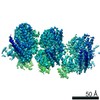
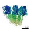
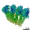
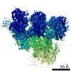
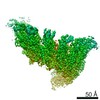
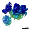
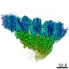
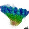
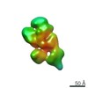
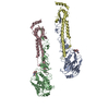
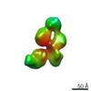
| Title | Structures of GCP2 and GCP3 in the native human gamma-tubulin ring complex | |||||||||||||||||||||||||||||||||
 Components Components |
| |||||||||||||||||||||||||||||||||
 Keywords Keywords | STRUCTURAL PROTEIN / GCP / GCP2 / GCP3 / gamma-TuSC / gTuSC / gamma-tubulin ring complex / gTuRC / g-TuRC / microtubule / microtubule nucleation / single particle cryo-EM structure | |||||||||||||||||||||||||||||||||
| Function / homology |  Function and homology information Function and homology informationpolar microtubule / gamma-tubulin complex / microtubule nucleation / gamma-tubulin binding / single fertilization / microtubule organizing center / cytoplasmic microtubule / spindle assembly / cytoplasmic microtubule organization / Recruitment of mitotic centrosome proteins and complexes ...polar microtubule / gamma-tubulin complex / microtubule nucleation / gamma-tubulin binding / single fertilization / microtubule organizing center / cytoplasmic microtubule / spindle assembly / cytoplasmic microtubule organization / Recruitment of mitotic centrosome proteins and complexes / Recruitment of NuMA to mitotic centrosomes / centriole / brain development / meiotic cell cycle / structural constituent of cytoskeleton / neuron migration / spindle / spindle pole / mitotic cell cycle / protein-containing complex assembly / ciliary basal body / centrosome / structural molecule activity / nucleoplasm / membrane / cytosol / cytoplasm Similarity search - Function | |||||||||||||||||||||||||||||||||
| Biological species |  Homo sapiens (human) Homo sapiens (human) | |||||||||||||||||||||||||||||||||
| Method | ELECTRON MICROSCOPY / single particle reconstruction / cryo EM / Resolution: 3.8 Å | |||||||||||||||||||||||||||||||||
 Authors Authors | Wieczorek, M. / Urnavicius, L. / Ti, S. / Molloy, K.R. / Chait, B.T. / Kapoor, T.M. | |||||||||||||||||||||||||||||||||
| Funding support |  United States, United States,  France, 2items France, 2items
| |||||||||||||||||||||||||||||||||
 Citation Citation | Journal: Cell / Year: 2020 Title: Asymmetric Molecular Architecture of the Human γ-Tubulin Ring Complex. Authors: Michal Wieczorek / Linas Urnavicius / Shih-Chieh Ti / Kelly R Molloy / Brian T Chait / Tarun M Kapoor / Abstract: The γ-tubulin ring complex (γ-TuRC) is an essential regulator of centrosomal and acentrosomal microtubule formation, yet its structure is not known. Here, we present a cryo-EM reconstruction of the ...The γ-tubulin ring complex (γ-TuRC) is an essential regulator of centrosomal and acentrosomal microtubule formation, yet its structure is not known. Here, we present a cryo-EM reconstruction of the native human γ-TuRC at ∼3.8 Å resolution, revealing an asymmetric, cone-shaped structure. Pseudo-atomic models indicate that GCP4, GCP5, and GCP6 form distinct Y-shaped assemblies that structurally mimic GCP2/GCP3 subcomplexes distal to the γ-TuRC "seam." We also identify an unanticipated structural bridge that includes an actin-like protein and spans the γ-TuRC lumen. Despite its asymmetric architecture, the γ-TuRC arranges γ-tubulins into a helical geometry poised to nucleate microtubules. Diversity in the γ-TuRC subunits introduces large (>100,000 Å) surfaces in the complex that allow for interactions with different regulatory factors. The observed compositional complexity of the γ-TuRC could self-regulate its assembly into a cone-shaped structure to control microtubule formation across diverse contexts, e.g., within biological condensates or alongside existing filaments. | |||||||||||||||||||||||||||||||||
| History |
|
- Structure visualization
| Movie |
Movie viewer |
|---|---|
| Structure viewer | Molecule: MolmilJmol/JSmol |
- Downloads & links
-Download
| PDBx/mmCIF format | 6v6b.cif.gz | 223.3 KB | Display | PDBx/mmCIF format |
|---|---|---|---|---|
| PDB format | pdb6v6b.ent.gz | 169.6 KB | Display | PDB format |
| PDBx/mmJSON format | 6v6b.json.gz | Tree view | PDBx/mmJSON format | |
| Others |  Other downloads Other downloads |
-Validation report
| Arichive directory | https://data.pdbj.org/pub/pdb/validation_reports/v6/6v6bftp://data.pdbj.org/pub/pdb/validation_reports/v6/6v6b | HTTPS FTP |
|---|
-Related structure data
| Related structure data | 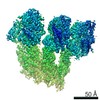 21067MC 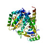 6v5vC 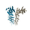 6v69C 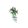 6v6cC 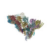 6v6sC M: map data used to model this data C: citing same article ( |
|---|---|
| Similar structure data |


-Links
 PDBj
PDBj
- Assembly
| Deposited unit | 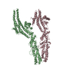
|
|---|---|
| 1 |
|
-Components
| #1: Protein | Mass: 103710.102 Da / Num. of mol.: 1 / Source method: isolated from a natural source / Source: (natural) Homo sapiens (human) / References: UniProt: Q96CW5 |
|---|---|
| #2: Protein | Mass: 105765.719 Da / Num. of mol.: 1 / Source method: isolated from a natural source / Source: (natural) Homo sapiens (human) / References: UniProt: Q9BSJ2 |
| Has protein modification | N |
-Experimental details
-Experiment
| Experiment | Method: ELECTRON MICROSCOPY |
|---|---|
| EM experiment | Aggregation state: PARTICLE / 3D reconstruction method: single particle reconstruction |
- Sample preparation
| Component | Name: Native human gamma-tubulin ring complex / Type: COMPLEX / Entity ID: all / Source: NATURAL |
|---|---|
| Molecular weight | Experimental value: NO |
| Source (natural) | Organism: Homo sapiens (human) |
| Buffer solution | pH: 7.5 |
| Specimen | Embedding applied: NO / Shadowing applied: NO / Staining applied: NO / Vitrification applied: YES |
| Vitrification | Cryogen name: ETHANE |
- Electron microscopy imaging
| Experimental equipment |  Model: Titan Krios / Image courtesy: FEI Company |
|---|---|
| Microscopy | Model: FEI TITAN KRIOS |
| Electron gun | Electron source:  FIELD EMISSION GUN / Accelerating voltage: 300 kV / Illumination mode: OTHER FIELD EMISSION GUN / Accelerating voltage: 300 kV / Illumination mode: OTHER |
| Electron lens | Mode: BRIGHT FIELD |
| Image recording | Electron dose: 45 e/Å2 / Film or detector model: GATAN K2 SUMMIT (4k x 4k) |
- Processing
| Software | Name: PHENIX / Version: 1.16_3549: / Classification: refinement | ||||||||||||||||||||||||
|---|---|---|---|---|---|---|---|---|---|---|---|---|---|---|---|---|---|---|---|---|---|---|---|---|---|
| EM software | Name: PHENIX / Category: model refinement | ||||||||||||||||||||||||
| CTF correction | Type: PHASE FLIPPING AND AMPLITUDE CORRECTION | ||||||||||||||||||||||||
| 3D reconstruction | Resolution: 3.8 Å / Resolution method: FSC 0.143 CUT-OFF / Num. of particles: 351714 / Symmetry type: POINT | ||||||||||||||||||||||||
| Refine LS restraints |
|
