 Movie
Movie Controller


+ Open data

- Basic information
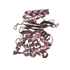
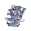
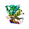
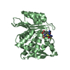
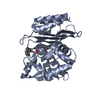
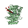
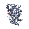
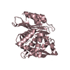
| Entry | Database: PDB / ID: 6v5e | ||||||
|---|---|---|---|---|---|---|---|
| Title | Crystal structure of CTX-M-14 P167S/D240G beta-lactamase | ||||||
 Components Components | Beta-lactamase | ||||||
 Keywords Keywords | HYDROLASE / drug resistance / protein evolution / conformational change / enzyme kinetics / enzyme catalysis / protein stability / protein-drug interaction | ||||||
| Function / homology |  Function and homology information Function and homology informationbeta-lactam antibiotic catabolic process / beta-lactamase activity / beta-lactamase / response to antibiotic / metal ion binding Similarity search - Function | ||||||
| Biological species |  | ||||||
| Method |  X-RAY DIFFRACTION / SYNCHROTRON / MOLECULAR REPLACEMENT / Resolution: 2.3 Å X-RAY DIFFRACTION / SYNCHROTRON / MOLECULAR REPLACEMENT / Resolution: 2.3 Å | ||||||
 Authors Authors | Brown, C.A. / Hu, L. / Sankaran, B. / Prasad, B.V.V. / Palzkill, T.G. | ||||||
| Funding support |  United States, 1items United States, 1items
| ||||||
 Citation Citation | Journal: J.Biol.Chem. / Year: 2020 Title: Antagonism between substitutions in beta-lactamase explains a path not taken in the evolution of bacterial drug resistance. Authors: Brown, C.A. / Hu, L. / Sun, Z. / Patel, M.P. / Singh, S. / Porter, J.R. / Sankaran, B. / Prasad, B.V.V. / Bowman, G.R. / Palzkill, T. | ||||||
| History |
|
- Structure visualization
| Structure viewer | Molecule: MolmilJmol/JSmol |
|---|
- Downloads & links
-Download
| PDBx/mmCIF format | 6v5e.cif.gz | 136.1 KB | Display | PDBx/mmCIF format |
|---|---|---|---|---|
| PDB format | pdb6v5e.ent.gz | 86.4 KB | Display | PDB format |
| PDBx/mmJSON format | 6v5e.json.gz | Tree view | PDBx/mmJSON format | |
| Others |  Other downloads Other downloads |
-Validation report
| Arichive directory | https://data.pdbj.org/pub/pdb/validation_reports/v5/6v5eftp://data.pdbj.org/pub/pdb/validation_reports/v5/6v5e | HTTPS FTP |
|---|
-Related structure data
| Related structure data | 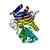 6v6gC 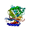 6v6pC 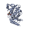 6v7tC 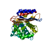 6v83C  6v8vC  1yltS S: Starting model for refinement C: citing same article ( |
|---|---|
| Similar structure data |


-Links
 PDBj
PDBj


- Assembly
| Deposited unit | 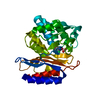
| ||||||||||||
|---|---|---|---|---|---|---|---|---|---|---|---|---|---|
| 1 |
| ||||||||||||
| Unit cell |
| ||||||||||||
| Components on special symmetry positions |
|
-Components
| #1: Protein | Mass: 27993.297 Da / Num. of mol.: 1 / Mutation: P167S, D240G Source method: isolated from a genetically manipulated source Source: (gene. exp.) Gene: blaCTX-M-14, beta-lactamase CTX-M-14, bla, bla CTX-M-14, bla-CTX-M-14a, blaCTX-M, blaCTX-M-14a, blaCTX-M-14b, blaCTX-M-14c, blaCTX-M-27b, blatoho-3, blaUOE-2, CTX-M-14, AM465_01285, AM465_ ...Gene: blaCTX-M-14, beta-lactamase CTX-M-14, bla, bla CTX-M-14, bla-CTX-M-14a, blaCTX-M, blaCTX-M-14a, blaCTX-M-14b, blaCTX-M-14c, blaCTX-M-27b, blatoho-3, blaUOE-2, CTX-M-14, AM465_01285, AM465_06510, AM465_23360, APT94_14605, BEN53_26220, BET08_34355, BJJ90_27545, BK334_27290, BOH76_00730, BON63_16015, BON66_01305, BON69_22545, BON71_04040, BON75_10525, BON76_14325, BON81_01055, BON83_15455, BON86_08515, BON91_02075, BON92_04750, BON94_23850, BON95_01680, BON96_03940, BXT93_06855, C5N07_28500, C5P43_21980, CDL37_21060, CR538_26855, CRT46_23505, DW236_20870, EIA08_25160, EIA21_26975, ELT23_05930, ETN48_p0088, FNJ69_13810, FQR64_24895, FTV90_03295, pCT_085, pHK01_011, RCS103_P0010, RCS30_P0082, RCS56_P0085, RCS60_P0031, RCS63_P0006, RCS65_P0008, RCS66_P0053, SAMEA4362930_00013, SAMEA4370290_00046 Production host: |
|---|---|
| #2: Water | ChemComp-HOH /  Mass: 18.015 Da / Num. of mol.: 124 / Source method: isolated from a natural source / Formula: H2O Mass: 18.015 Da / Num. of mol.: 124 / Source method: isolated from a natural source / Formula: H2O |
| Has ligand of interest | N |
-Experimental details
-Experiment
| Experiment | Method: X-RAY DIFFRACTION / Number of used crystals: 1 |
|---|
- Sample preparation
| Crystal | Density Matthews: 2.09 Å3/Da / Density % sol: 41.11 % |
|---|---|
| Crystal grow | Temperature: 293 K / Method: vapor diffusion, hanging drop / pH: 4 / Details: 0.1 M MIB Buffer pH 4.0, 25% (w/v) PEG 1500 |
-Data collection
| Diffraction | Mean temperature: 100 K / Serial crystal experiment: N |
|---|---|
| Diffraction source | Source: SYNCHROTRON / Site: ALS / Beamline: 5.0.2 / Wavelength: 0.97741 Å |
| Detector | Type: DECTRIS PILATUS 6M / Detector: PIXEL / Date: Mar 18, 2016 |
| Radiation | Protocol: SINGLE WAVELENGTH / Monochromatic (M) / Laue (L): M / Scattering type: x-ray |
| Radiation wavelength | Wavelength: 0.97741 Å / Relative weight: 1 |
| Reflection | Resolution: 2.3→41.7 Å / Num. obs: 10104 / % possible obs: 88.28 % / Redundancy: 7 % / Biso Wilson estimate: 29.57 Å2 / Rmerge(I) obs: 0.084 / Rpim(I) all: 0.033 / Rrim(I) all: 0.091 / Net I/σ(I): 17.4 |
| Reflection shell | Resolution: 2.3→2.38 Å / Rmerge(I) obs: 0.121 / Num. unique obs: 880 / CC1/2: 0.978 / Rpim(I) all: 0.046 / Rrim(I) all: 0.13 |
- Processing
| Software |
| |||||||||||||||||||||||||||||||||||||||||||||||||||||||||||||||||||||||||||||||||||||||||||||||||||||||||||||||||||||||||||||
|---|---|---|---|---|---|---|---|---|---|---|---|---|---|---|---|---|---|---|---|---|---|---|---|---|---|---|---|---|---|---|---|---|---|---|---|---|---|---|---|---|---|---|---|---|---|---|---|---|---|---|---|---|---|---|---|---|---|---|---|---|---|---|---|---|---|---|---|---|---|---|---|---|---|---|---|---|---|---|---|---|---|---|---|---|---|---|---|---|---|---|---|---|---|---|---|---|---|---|---|---|---|---|---|---|---|---|---|---|---|---|---|---|---|---|---|---|---|---|---|---|---|---|---|---|---|---|
| Refinement | Method to determine structure: MOLECULAR REPLACEMENT Starting model: 1YLT Resolution: 2.3→41.7 Å / SU ML: 0.2733 / Cross valid method: FREE R-VALUE / σ(F): 1.34 / Phase error: 21.9359 Stereochemistry target values: GeoStd + Monomer Library + CDL v1.2
| |||||||||||||||||||||||||||||||||||||||||||||||||||||||||||||||||||||||||||||||||||||||||||||||||||||||||||||||||||||||||||||
| Solvent computation | Shrinkage radii: 0.9 Å / VDW probe radii: 1.11 Å / Solvent model: FLAT BULK SOLVENT MODEL | |||||||||||||||||||||||||||||||||||||||||||||||||||||||||||||||||||||||||||||||||||||||||||||||||||||||||||||||||||||||||||||
| Displacement parameters | Biso mean: 31 Å2 | |||||||||||||||||||||||||||||||||||||||||||||||||||||||||||||||||||||||||||||||||||||||||||||||||||||||||||||||||||||||||||||
| Refinement step | Cycle: LAST / Resolution: 2.3→41.7 Å
| |||||||||||||||||||||||||||||||||||||||||||||||||||||||||||||||||||||||||||||||||||||||||||||||||||||||||||||||||||||||||||||
| Refine LS restraints |
| |||||||||||||||||||||||||||||||||||||||||||||||||||||||||||||||||||||||||||||||||||||||||||||||||||||||||||||||||||||||||||||
| LS refinement shell |
| |||||||||||||||||||||||||||||||||||||||||||||||||||||||||||||||||||||||||||||||||||||||||||||||||||||||||||||||||||||||||||||
| Refinement TLS params. | Method: refined / Refine-ID: X-RAY DIFFRACTION
| |||||||||||||||||||||||||||||||||||||||||||||||||||||||||||||||||||||||||||||||||||||||||||||||||||||||||||||||||||||||||||||
| Refinement TLS group |
|
