 Movie
Movie Controller


+ Open data

- Basic information
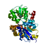
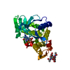
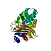
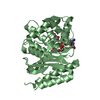
| Entry | Database: PDB / ID: 1ylt | |||||||||
|---|---|---|---|---|---|---|---|---|---|---|
| Title | Atomic resolution structure of CTX-M-14 beta-lactamase | |||||||||
 Components Components | beta-lactamase CTX-M-14 | |||||||||
 Keywords Keywords | HYDROLASE / CTX-M / beta-lactamase / anisotropy / extended-spectrum | |||||||||
| Function / homology |  Function and homology information Function and homology informationbeta-lactam antibiotic catabolic process / beta-lactamase activity / beta-lactamase / response to antibiotic / metal ion binding Similarity search - Function | |||||||||
| Biological species |  | |||||||||
| Method |  X-RAY DIFFRACTION / SYNCHROTRON / AB INITIO / Resolution: 1.1 Å X-RAY DIFFRACTION / SYNCHROTRON / AB INITIO / Resolution: 1.1 Å | |||||||||
 Authors Authors | Chen, Y. / Delmas, J. / Sirot, J. / Shoichet, B. / Bonnet, R. | |||||||||
 Citation Citation | Journal: J.Mol.Biol. / Year: 2005 Title: Atomic Resolution Structures of CTX-M beta-Lactamases: Extended Spectrum Activities from Increased Mobility and Decreased Stability. Authors: Chen, Y. / Delmas, J. / Sirot, J. / Shoichet, B. / Bonnet, R. | |||||||||
| History |
|
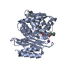
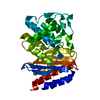
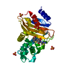
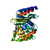
- Structure visualization
| Structure viewer | Molecule: MolmilJmol/JSmol |
|---|
- Downloads & links
-Download
| PDBx/mmCIF format | 1ylt.cif.gz | 139.4 KB | Display | PDBx/mmCIF format |
|---|---|---|---|---|
| PDB format | pdb1ylt.ent.gz | 106.5 KB | Display | PDB format |
| PDBx/mmJSON format | 1ylt.json.gz | Tree view | PDBx/mmJSON format | |
| Others |  Other downloads Other downloads |
-Validation report
| Arichive directory | https://data.pdbj.org/pub/pdb/validation_reports/yl/1yltftp://data.pdbj.org/pub/pdb/validation_reports/yl/1ylt | HTTPS FTP |
|---|
-Related structure data
| Related structure data |  1yljSC 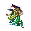 1ylpC 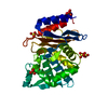 1ylwC S: Starting model for refinement C: citing same article ( |
|---|---|
| Similar structure data |


-Links
 PDBj
PDBj


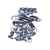
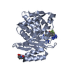
- Assembly
| Deposited unit | 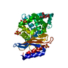
| ||||||||
|---|---|---|---|---|---|---|---|---|---|
| 1 |
| ||||||||
| Unit cell |
|
-Components
| #1: Protein | Mass: 28000.547 Da / Num. of mol.: 1 / Mutation: A231V Source method: isolated from a genetically manipulated source Source: (gene. exp.) Keywords: CTX-M-14 beta-lactamase / References: UniProt: Q9L5C7, beta-lactamase | ||
|---|---|---|---|
| #2: Polysaccharide | beta-D-fructofuranose-(2-1)-alpha-D-glucopyranose / sucrose  Source method: isolated from a genetically manipulated source Details: oligosaccharide with reducing-end-to-reducing-end glycosidic bond References: sucrose | ||
| #3: Chemical | ChemComp-SO4 /   Mass: 96.063 Da / Num. of mol.: 8 / Source method: obtained synthetically / Formula: SO4 Mass: 96.063 Da / Num. of mol.: 8 / Source method: obtained synthetically / Formula: SO4#4: Water | ChemComp-HOH / |  Mass: 18.015 Da / Num. of mol.: 427 / Source method: isolated from a natural source / Formula: H2O Mass: 18.015 Da / Num. of mol.: 427 / Source method: isolated from a natural source / Formula: H2O |
-Experimental details
-Experiment
| Experiment | Method: X-RAY DIFFRACTION / Number of used crystals: 1 |
|---|
- Sample preparation
| Crystal | Density Matthews: 1.72 Å3/Da / Density % sol: 28.4 % |
|---|---|
| Crystal grow | Temperature: 293 K / pH: 4.5 Details: ammonium sulfate, sodium acetate, pH 4.5, VAPOR DIFFUSION, HANGING DROP, temperature 293K, pH 4.50 |
-Data collection
| Diffraction | Mean temperature: 100 K |
|---|---|
| Diffraction source | Source: SYNCHROTRON / Site: ALS  / Beamline: 8.3.1 / Wavelength: 1.033 / Beamline: 8.3.1 / Wavelength: 1.033 |
| Detector | Type: ADSC QUANTUM 4 / Detector: CCD / Date: Feb 12, 2004 / Details: MIRRORS |
| Radiation | Monochromator: DOUBLE CRYSTAL / Protocol: SINGLE WAVELENGTH / Monochromatic (M) / Laue (L): M / Scattering type: x-ray |
| Radiation wavelength | Wavelength: 1.033 Å / Relative weight: 1 |
| Reflection | Resolution: 1.1→22 Å / Num. obs: 89046 / % possible obs: 96.9 % / Observed criterion σ(I): -3 / Redundancy: 6.23 % / Rmerge(I) obs: 0.037 / Net I/σ(I): 16.2 |
| Reflection shell | Resolution: 1.1→1.14 Å / Rmerge(I) obs: 0.276 / Mean I/σ(I) obs: 2.02 / % possible all: 76.4 |
- Processing
| Software |
| |||||||||||||||||||||||||||||||||
|---|---|---|---|---|---|---|---|---|---|---|---|---|---|---|---|---|---|---|---|---|---|---|---|---|---|---|---|---|---|---|---|---|---|---|
| Refinement | Method to determine structure: AB INITIO Starting model: PDB ENTRY 1YLJ Resolution: 1.1→22 Å / Num. parameters: 23478 / Num. restraintsaints: 32095 / Cross valid method: FREE R / σ(F): 0 / Stereochemistry target values: ENGH AND HUBER Details: ANISOTROPIC REFINEMENT. RIDING HYDROGENS INCLUDED IN REFINEMENT. WATER MOLECULES IN CLOSE CONTACT HAVE A COMBINED OCCUPANCY OF 1.
| |||||||||||||||||||||||||||||||||
| Refine analyze | Num. disordered residues: 122 / Occupancy sum hydrogen: 1886 / Occupancy sum non hydrogen: 2391.66 | |||||||||||||||||||||||||||||||||
| Refinement step | Cycle: LAST / Resolution: 1.1→22 Å
| |||||||||||||||||||||||||||||||||
| Refine LS restraints |
|
