 Movie
Movie Controller

[English] 日本語
 Yorodumi
- PDB-1yms: X-ray crystallographic structure of CTX-M-9 beta-lactamase comple... -
+ Open data

- Basic information
| Entry | Database: PDB / ID: 1yms | ||||||
|---|---|---|---|---|---|---|---|
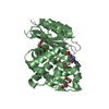
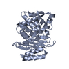
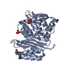
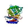
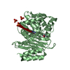
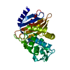
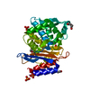
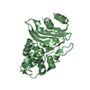
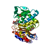
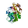
| Title | X-ray crystallographic structure of CTX-M-9 beta-lactamase complexed with nafcinin-like boronic acid inhibitor | ||||||
 Components Components | beta-lactamase CTX-M-9 | ||||||
 Keywords Keywords | HYDROLASE / CTX-M / beta-lactamase / transition state / acylation | ||||||
| Function / homology |  Function and homology information Function and homology informationbeta-lactam antibiotic catabolic process / beta-lactamase activity / beta-lactamase / response to antibiotic Similarity search - Function | ||||||
| Biological species |  | ||||||
| Method |  X-RAY DIFFRACTION / SYNCHROTRON / MOLECULAR REPLACEMENT / Resolution: 1.6 Å X-RAY DIFFRACTION / SYNCHROTRON / MOLECULAR REPLACEMENT / Resolution: 1.6 Å | ||||||
 Authors Authors | Chen, Y. / Shoichet, B. / Bonnet, R. | ||||||
 Citation Citation | Journal: J.Am.Chem.Soc. / Year: 2005 Title: Structure, Function, and Inhibition along the Reaction Coordinate of CTX-M beta-Lactamases. Authors: Chen, Y. / Shoichet, B. / Bonnet, R. | ||||||
| History |
|
- Structure visualization
| Structure viewer | Molecule: MolmilJmol/JSmol |
|---|
- Downloads & links
-Download
| PDBx/mmCIF format | 1yms.cif.gz | 129.1 KB | Display | PDBx/mmCIF format |
|---|---|---|---|---|
| PDB format | pdb1yms.ent.gz | 98.9 KB | Display | PDB format |
| PDBx/mmJSON format | 1yms.json.gz | Tree view | PDBx/mmJSON format | |
| Others |  Other downloads Other downloads |
-Validation report
| Arichive directory | https://data.pdbj.org/pub/pdb/validation_reports/ym/1ymsftp://data.pdbj.org/pub/pdb/validation_reports/ym/1yms | HTTPS FTP |
|---|
-Related structure data
| Related structure data |  1ylyC 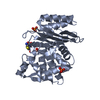 1ylzC 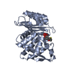 1ym1C 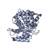 1ymxC  1yljS S: Starting model for refinement C: citing same article ( |
|---|---|
| Similar structure data |


-Links
 PDBj
PDBj


- Assembly
| Deposited unit | 
| ||||||||
|---|---|---|---|---|---|---|---|---|---|
| 1 | 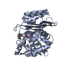
| ||||||||
| 2 | 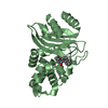
| ||||||||
| Unit cell |
|
-Components
| #1: Protein | Mass: 27972.494 Da / Num. of mol.: 2 Source method: isolated from a genetically manipulated source Source: (gene. exp.) Keywords: CTX-M-9 beta-lactamase, V231A / References: UniProt: Q9L5C8, beta-lactamase#2: Chemical |   Mass: 273.092 Da / Num. of mol.: 2 / Source method: obtained synthetically / Formula: C14H16BNO4 Mass: 273.092 Da / Num. of mol.: 2 / Source method: obtained synthetically / Formula: C14H16BNO4#3: Water | ChemComp-HOH / |  Mass: 18.015 Da / Num. of mol.: 804 / Source method: isolated from a natural source / Formula: H2O Mass: 18.015 Da / Num. of mol.: 804 / Source method: isolated from a natural source / Formula: H2OHas protein modification | Y | |
|---|
-Experimental details
-Experiment
| Experiment | Method: X-RAY DIFFRACTION / Number of used crystals: 1 |
|---|
- Sample preparation
| Crystal | Density Matthews: 2.02 Å3/Da / Density % sol: 38.76 % |
|---|---|
| Crystal grow | Temperature: 293 K / Method: vapor diffusion, hanging drop / pH: 8.8 Details: potassium phosphate, pH 8.8, VAPOR DIFFUSION, HANGING DROP, temperature 293K |
-Data collection
| Diffraction | Mean temperature: 100 K |
|---|---|
| Diffraction source | Source: SYNCHROTRON / Site: ALS  / Beamline: 8.3.1 / Wavelength: 1.11587 / Wavelength: 1.11587 Å / Beamline: 8.3.1 / Wavelength: 1.11587 / Wavelength: 1.11587 Å |
| Detector | Type: ADSC QUANTUM 4 / Detector: CCD / Date: Jul 17, 2004 / Details: mirrors |
| Radiation | Monochromator: Double crystal / Protocol: SINGLE WAVELENGTH / Monochromatic (M) / Laue (L): M / Scattering type: x-ray |
| Radiation wavelength | Wavelength: 1.11587 Å / Relative weight: 1 |
| Reflection | Resolution: 1.6→22 Å / Num. all: 58748 / Num. obs: 57897 / % possible obs: 98.6 % / Observed criterion σ(F): 0 / Observed criterion σ(I): -3 / Redundancy: 5.22 % / Biso Wilson estimate: 10.8 Å2 / Rmerge(I) obs: 0.069 / Net I/σ(I): 11 |
| Reflection shell | Resolution: 1.6→1.66 Å / Rmerge(I) obs: 0.347 / Mean I/σ(I) obs: 2.48 / % possible all: 96.7 |
- Processing
| Software |
| ||||||||||||||||||||||||||||||||||||
|---|---|---|---|---|---|---|---|---|---|---|---|---|---|---|---|---|---|---|---|---|---|---|---|---|---|---|---|---|---|---|---|---|---|---|---|---|---|
| Refinement | Method to determine structure: MOLECULAR REPLACEMENT Starting model: 1YLJ Resolution: 1.6→19.75 Å / Rfactor Rfree error: 0.004 / Data cutoff high absF: 81987.04 / Data cutoff low absF: 0 / Isotropic thermal model: RESTRAINED / Cross valid method: THROUGHOUT / σ(F): 0 / Stereochemistry target values: Engh & Huber Details: ONLY 5 ATOMS WERE MODELED FOR A25 GLN with the side chain truncated after CB
| ||||||||||||||||||||||||||||||||||||
| Solvent computation | Solvent model: FLAT MODEL / Bsol: 41.3739 Å2 / ksol: 0.36281 e/Å3 | ||||||||||||||||||||||||||||||||||||
| Displacement parameters | Biso mean: 10.8 Å2
| ||||||||||||||||||||||||||||||||||||
| Refine analyze |
| ||||||||||||||||||||||||||||||||||||
| Refinement step | Cycle: LAST / Resolution: 1.6→19.75 Å
| ||||||||||||||||||||||||||||||||||||
| Refine LS restraints |
| ||||||||||||||||||||||||||||||||||||
| LS refinement shell | Resolution: 1.6→1.7 Å / Rfactor Rfree error: 0.012 / Total num. of bins used: 6
| ||||||||||||||||||||||||||||||||||||
| Xplor file |
|
