 Movie
Movie Controller


+ Open data

- Basic information
| Entry | Database: PDB / ID: 1dy6 | ||||||
|---|---|---|---|---|---|---|---|
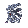
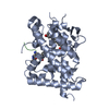
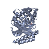
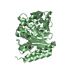
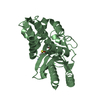
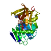
| Title | Structure of the imipenem-hydrolyzing beta-lactamase SME-1 | ||||||
 Components Components | CARBAPENEM-HYDROLYSING BETA-LACTAMASE SME-1 | ||||||
 Keywords Keywords | HYDROLASE / LACTAMASE / ANTIBIOTIC / CARBAPENEM / IMIPENEM | ||||||
| Function / homology |  Function and homology information Function and homology informationbeta-lactam antibiotic catabolic process / beta-lactamase activity / beta-lactamase / response to antibiotic Similarity search - Function | ||||||
| Biological species |  SERRATIA MARCESCENS (bacteria) SERRATIA MARCESCENS (bacteria) | ||||||
| Method |  X-RAY DIFFRACTION / MOLECULAR REPLACEMENT / Resolution: 2.13 Å X-RAY DIFFRACTION / MOLECULAR REPLACEMENT / Resolution: 2.13 Å | ||||||
 Authors Authors | Sougakoff, W. / L'Hermite, G. / Billy, I. / Guillet, V. / Naas, T. / Nordman, P. / Jarlier, V. / Delettre, J. | ||||||
 Citation Citation | Journal: Acta Crystallogr.,Sect.D / Year: 2002 Title: Structure of the Imipenem-Hydrolyzing Class a Beta-Lactamase Sme-1 from Serratia Marcescens. Authors: Sougakoff, W. / L'Hermite, G. / Billy, I. / Pernot, L. / Guillet, V. / Naas, T. / Nordmann, P. / Jarlier, V. / Delettre, J. #1: Journal: J.Struct.Biol. / Year: 1996 Title: Purification, Crystallization, and Preliminary X-Ray Diffraction Analysis of the Carbapenem-Hydrolyzing Class a Beta-Lactamase Sme-1 from Serratia Marcescens Authors: Sougakoff, W. / Jarlier, V. / Delettre, J. / Colloc'H, N. / L'Hermite, G. / Nordmann, P. / Naas, T. | ||||||
| History |
|
- Structure visualization
| Structure viewer | Molecule: MolmilJmol/JSmol |
|---|
- Downloads & links
-Download
| PDBx/mmCIF format | 1dy6.cif.gz | 120.7 KB | Display | PDBx/mmCIF format |
|---|---|---|---|---|
| PDB format | pdb1dy6.ent.gz | 93.9 KB | Display | PDB format |
| PDBx/mmJSON format | 1dy6.json.gz | Tree view | PDBx/mmJSON format | |
| Others |  Other downloads Other downloads |
-Validation report
| Arichive directory | https://data.pdbj.org/pub/pdb/validation_reports/dy/1dy6ftp://data.pdbj.org/pub/pdb/validation_reports/dy/1dy6 | HTTPS FTP |
|---|
-Related structure data
| Related structure data | 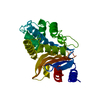 1bueS S: Starting model for refinement |
|---|---|
| Similar structure data |


-Links
 PDBj
PDBj


- Assembly
| Deposited unit | 
| ||||||||
|---|---|---|---|---|---|---|---|---|---|
| 1 | 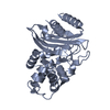
| ||||||||
| 2 | 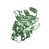
| ||||||||
| Unit cell |
| ||||||||
| Noncrystallographic symmetry (NCS) | NCS oper: (Code: given Matrix: (0.986365, 0.004914, 0.164497), Vector: Details | BIOLOGICAL UNIT: MONOMER | |
-Components
| #1: Protein | Mass: 29386.180 Da / Num. of mol.: 2 Source method: isolated from a genetically manipulated source Details: APO FORM / Source: (gene. exp.) SERRATIA MARCESCENS (bacteria) / Strain: S6 / Plasmid: PPTN103 / Gene (production host): BLASME / Production host: #2: Water | ChemComp-HOH / |  Mass: 18.015 Da / Num. of mol.: 397 / Source method: isolated from a natural source / Formula: H2O Mass: 18.015 Da / Num. of mol.: 397 / Source method: isolated from a natural source / Formula: H2OHas protein modification | Y | Sequence details | SEQUENCE DESCRIBED IN NAAS T., VANDEL L., SOUGAKOFF W., LIVERMORE D.M., NORDMANN P. ANTIMICROB. ...SEQUENCE DESCRIBED IN NAAS T., VANDEL L., SOUGAKOFF W., LIVERMORE D.M., NORDMANN P. ANTIMICROB | |
|---|
-Experimental details
-Experiment
| Experiment | Method: X-RAY DIFFRACTION / Number of used crystals: 1 |
|---|
- Sample preparation
| Crystal | Density Matthews: 2.26 Å3/Da / Density % sol: 40 % | ||||||||||||||||||||
|---|---|---|---|---|---|---|---|---|---|---|---|---|---|---|---|---|---|---|---|---|---|
| Crystal grow | pH: 8.5 Details: PROTEIN WAS CRYSTALLIZED FROM 17% PEG 4000, 0.2 M AMMONIUM ACETATE, 0.1 M TRIS-HCL, PH 8.5 | ||||||||||||||||||||
| Crystal grow | *PLUS Method: vapor diffusion, hanging drop | ||||||||||||||||||||
| Components of the solutions | *PLUS
|
-Data collection
| Diffraction | Mean temperature: 291 K |
|---|---|
| Diffraction source | Source: ROTATING ANODE / Type: ENRAF-NONIUS FR571 / Wavelength: 1.5418 |
| Detector | Type: ENRAF-NONIUS FAST / Detector: DIFFRACTOMETER |
| Radiation | Monochromator: NI FILTER / Protocol: SINGLE WAVELENGTH / Monochromatic (M) / Laue (L): M / Scattering type: x-ray |
| Radiation wavelength | Wavelength: 1.5418 Å / Relative weight: 1 |
| Reflection | Resolution: 2.13→20 Å / Num. obs: 24447 / % possible obs: 82 % / Redundancy: 2.3 % / Rsym value: 0.049 |
| Reflection | *PLUS % possible obs: 82 % / Num. measured all: 55772 / Rmerge(I) obs: 0.049 |
| Reflection shell | *PLUS Highest resolution: 2.13 Å / Lowest resolution: 2.28 Å / % possible obs: 30.1 % / Redundancy: 2.1 % / Num. unique obs: 1601 / Num. measured obs: 3390 / Rmerge(I) obs: 0.216 |
- Processing
| Software |
| ||||||||||||||||||||||||||||||||||||||||||||||||||||||||||||
|---|---|---|---|---|---|---|---|---|---|---|---|---|---|---|---|---|---|---|---|---|---|---|---|---|---|---|---|---|---|---|---|---|---|---|---|---|---|---|---|---|---|---|---|---|---|---|---|---|---|---|---|---|---|---|---|---|---|---|---|---|---|
| Refinement | Method to determine structure: MOLECULAR REPLACEMENT Starting model: MODIFIED NMC-A BETA-LACTAMASE (1BUE) SOFTWARE USED: AMORE Resolution: 2.13→10 Å / Rfactor Rfree error: 0.005 / Data cutoff low absF: 0.001 / Cross valid method: THROUGHOUT / σ(F): 0 Details: A FLAT TWO-LOBE ELECTRON DENSITY IS LOCATED IN A AND IN B BETWEEN SER130, THR235 AND SER237. IT HAS BEEN ASSOCIATED WITH A MODEL OF TWO WATER MOLECULES, 111 AND 186 IN MOLECULE A AND 89 AND 143 IN MOLECULE B.
| ||||||||||||||||||||||||||||||||||||||||||||||||||||||||||||
| Displacement parameters | Biso mean: 19.2 Å2 | ||||||||||||||||||||||||||||||||||||||||||||||||||||||||||||
| Refinement step | Cycle: LAST / Resolution: 2.13→10 Å
| ||||||||||||||||||||||||||||||||||||||||||||||||||||||||||||
| Refine LS restraints |
|
