 Movie
Movie Controller


+ Open data

- Basic information
| Entry | Database: PDB / ID: 1bue | ||||||
|---|---|---|---|---|---|---|---|
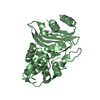
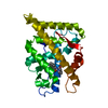
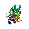
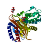
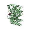
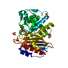
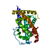
| Title | NMC-A CARBAPENEMASE FROM ENTEROBACTER CLOACAE | ||||||
 Components Components | PROTEIN (IMIPENEM-HYDROLYSING BETA-LACTAMASE) | ||||||
 Keywords Keywords | HYDROLASE / ANTIBIOTIC RESISTANCE / CLASS A CARBAPENEMASE | ||||||
| Function / homology |  Function and homology information Function and homology informationbeta-lactam antibiotic catabolic process / beta-lactamase activity / beta-lactamase / response to antibiotic Similarity search - Function | ||||||
| Biological species |  Enterobacter cloacae (bacteria) Enterobacter cloacae (bacteria) | ||||||
| Method |  X-RAY DIFFRACTION / SYNCHROTRON / SIRAS / Resolution: 1.64 Å X-RAY DIFFRACTION / SYNCHROTRON / SIRAS / Resolution: 1.64 Å | ||||||
 Authors Authors | Swaren, P. / Maveyraud, L. / Cabantous, S. / Pedelacq, J.D. / Mourey, L. / Frere, J.M. / Samama, J.P. | ||||||
 Citation Citation | Journal: J.Biol.Chem. / Year: 1998 Title: X-ray analysis of the NMC-A beta-lactamase at 1.64-A resolution, a class A carbapenemase with broad substrate specificity. Authors: Swaren, P. / Maveyraud, L. / Raquet, X. / Cabantous, S. / Duez, C. / Pedelacq, J.D. / Mariotte-Boyer, S. / Mourey, L. / Labia, R. / Nicolas-Chanoine, M.H. / Nordmann, P. / Frere, J.M. / Samama, J.P. #1: Journal: FEMS Microbiol.Lett. / Year: 1996Title: A Kinetic Study of NMC-A Beta-Lactamase, an Ambler Class A Carbapenemase Also Hydrolyzing Cephamycins Authors: Mariotte-Boyer, S. / Nicolas-Chanoine, M.H. / Labia, R. #2: Journal: Proc.Natl.Acad.Sci.USA / Year: 1994Title: Analysis of a Carbapenem-Hydrolyzing Class A Beta-Lactamase from Enterobacter Cloacae and of its Lysr-Type Regulatory Protein Authors: Naas, T. / Nordmann, P. | ||||||
| History |
|
- Structure visualization
| Structure viewer | Molecule: MolmilJmol/JSmol |
|---|
- Downloads & links
-Download
| PDBx/mmCIF format | 1bue.cif.gz | 65.2 KB | Display | PDBx/mmCIF format |
|---|---|---|---|---|
| PDB format | pdb1bue.ent.gz | 46.5 KB | Display | PDB format |
| PDBx/mmJSON format | 1bue.json.gz | Tree view | PDBx/mmJSON format | |
| Others |  Other downloads Other downloads |
-Validation report
| Arichive directory | https://data.pdbj.org/pub/pdb/validation_reports/bu/1bueftp://data.pdbj.org/pub/pdb/validation_reports/bu/1bue | HTTPS FTP |
|---|
-Related structure data
| Similar structure data |
|---|


-Links
 PDBj
PDBj


- Assembly
| Deposited unit | 
| ||||||||
|---|---|---|---|---|---|---|---|---|---|
| 1 |
| ||||||||
| Unit cell |
|
-Components
| #1: Protein | Mass: 29137.596 Da / Num. of mol.: 1 Source method: isolated from a genetically manipulated source Source: (gene. exp.) Enterobacter cloacae (bacteria) / Strain: NOR-1 / Production host: |
|---|---|
| #2: Water | ChemComp-HOH /  Mass: 18.015 Da / Num. of mol.: 115 / Source method: isolated from a natural source / Formula: H2O Mass: 18.015 Da / Num. of mol.: 115 / Source method: isolated from a natural source / Formula: H2O |
| Has protein modification | Y |
-Experimental details
-Experiment
| Experiment | Method: X-RAY DIFFRACTION / Number of used crystals: 2 |
|---|
- Sample preparation
| Crystal | Density Matthews: 2.4 Å3/Da / Density % sol: 45.6 % Description: ONE CRYSTAL WAS USED FOR NATIVE LOW RESOLUTION DATA TO 2.9 ANGSTROMS RESOLUTION, AND A SECOND CRYSTAL WAS USED FOR DATA TO 1.64 ANGSTROMS RESOLUTION. | |||||||||||||||||||||||||||||||||||
|---|---|---|---|---|---|---|---|---|---|---|---|---|---|---|---|---|---|---|---|---|---|---|---|---|---|---|---|---|---|---|---|---|---|---|---|---|
| Crystal grow | pH: 5.25 Details: INITIAL PROTEIN CONCENTRATION WAS 2.0 G/L EQUILIBRATED AGAINST 0.200 M MES PH 5.25, 20% (W/V)PEG 1500, 6% (V/V) N-PROPANOL AT 295K. | |||||||||||||||||||||||||||||||||||
| Crystal | *PLUS | |||||||||||||||||||||||||||||||||||
| Crystal grow | *PLUS Temperature: 22 ℃ / Method: vapor diffusion, hanging drop | |||||||||||||||||||||||||||||||||||
| Components of the solutions | *PLUS
|
-Data collection
| Diffraction | Mean temperature: 277 K |
|---|---|
| Diffraction source | Source: SYNCHROTRON / Site: LURE  / Beamline: DW32 / Wavelength: 0.975 / Beamline: DW32 / Wavelength: 0.975 |
| Detector | Type: MARRESEARCH / Detector: IMAGE PLATE / Date: May 7, 1996 |
| Radiation | Protocol: SINGLE WAVELENGTH / Monochromatic (M) / Laue (L): M / Scattering type: x-ray |
| Radiation wavelength | Wavelength: 0.975 Å / Relative weight: 1 |
| Reflection | Resolution: 1.64→31 Å / Num. obs: 32762 / % possible obs: 93.3 % / Redundancy: 4.1 % / Biso Wilson estimate: 13.6 Å2 / Rmerge(I) obs: 0.045 / Net I/σ(I): 33.3 |
| Reflection shell | Resolution: 1.64→1.68 Å / Redundancy: 2.3 % / Mean I/σ(I) obs: 7.1 / Rsym value: 0.192 / % possible all: 68 |
| Reflection | *PLUS Num. measured all: 111487 |
- Processing
| Software |
| ||||||||||||||||||||||||||||||||||||||||||||||||||||||||||||
|---|---|---|---|---|---|---|---|---|---|---|---|---|---|---|---|---|---|---|---|---|---|---|---|---|---|---|---|---|---|---|---|---|---|---|---|---|---|---|---|---|---|---|---|---|---|---|---|---|---|---|---|---|---|---|---|---|---|---|---|---|---|
| Refinement | Method to determine structure: SIRAS Starting model: NOT APPLICABLE Resolution: 1.64→31 Å / Cross valid method: THROUGHOUT / σ(F): 0 Details: BULK SOLVENT CORRECTION USED WITH A DENSITY OF 0.34 E-/A**3, SOLVENT RADIUS OF 0.25 ANGSTROMS AND B FACTORS OF 50 A**2 RESIDUES 30 AND 63 HAVE ALTERNATE CONFORMATIONS. RESIDUE 140A WAS INSERTED.
| ||||||||||||||||||||||||||||||||||||||||||||||||||||||||||||
| Displacement parameters | Biso mean: 13.3 Å2 | ||||||||||||||||||||||||||||||||||||||||||||||||||||||||||||
| Refine analyze | Luzzati coordinate error obs: 0.18 Å / Luzzati d res low obs: 31 Å / Luzzati sigma a obs: 0.16 Å | ||||||||||||||||||||||||||||||||||||||||||||||||||||||||||||
| Refinement step | Cycle: LAST / Resolution: 1.64→31 Å
| ||||||||||||||||||||||||||||||||||||||||||||||||||||||||||||
| Refine LS restraints |
| ||||||||||||||||||||||||||||||||||||||||||||||||||||||||||||
| Refine LS restraints NCS | NCS model details: NOT APPLICABLE | ||||||||||||||||||||||||||||||||||||||||||||||||||||||||||||
| LS refinement shell | Resolution: 1.64→1.71 Å / Total num. of bins used: 8 / % reflection obs: 74.3 % | ||||||||||||||||||||||||||||||||||||||||||||||||||||||||||||
| Software | *PLUS Name: X-PLOR / Version: 3.1 / Classification: refinement | ||||||||||||||||||||||||||||||||||||||||||||||||||||||||||||
| Refinement | *PLUS σ(F): 0 / % reflection Rfree: 5 % | ||||||||||||||||||||||||||||||||||||||||||||||||||||||||||||
| Solvent computation | *PLUS | ||||||||||||||||||||||||||||||||||||||||||||||||||||||||||||
| Displacement parameters | *PLUS Biso mean: 13.3 Å2 | ||||||||||||||||||||||||||||||||||||||||||||||||||||||||||||
| Refine LS restraints | *PLUS
|
