 Movie
Movie Controller


+ Open data

- Basic information
| Entry | Database: PDB / ID: 3hre | ||||||
|---|---|---|---|---|---|---|---|
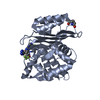
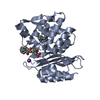
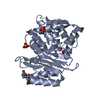
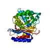
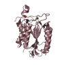
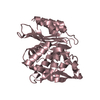
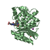
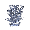
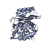
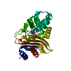
| Title | X-ray crystallographic structure of CTX-M-9 S70G | ||||||
 Components Components | CTX-M-9 extended-spectrum beta-lactamase | ||||||
 Keywords Keywords | HYDROLASE / beta-lactamase / BLSE / CTX-M-9 / Antibiotic resistance | ||||||
| Function / homology |  Function and homology information Function and homology informationbeta-lactam antibiotic catabolic process / beta-lactamase activity / beta-lactamase / response to antibiotic Similarity search - Function | ||||||
| Biological species |  | ||||||
| Method |  X-RAY DIFFRACTION / SYNCHROTRON / MOLECULAR REPLACEMENT / molecular replacement / Resolution: 1.45 Å X-RAY DIFFRACTION / SYNCHROTRON / MOLECULAR REPLACEMENT / molecular replacement / Resolution: 1.45 Å | ||||||
 Authors Authors | Delmas, J. / Leyssene, D. / Dubois, D. / Vazeille, E. / Robin, F. / Bonnet, R. | ||||||
 Citation Citation | Journal: J.Mol.Biol. / Year: 2010 Title: Structural insights into substrate recognition and product expulsion in CTX-M enzymes. Authors: Delmas, J. / Leyssene, D. / Dubois, D. / Birck, C. / Vazeille, E. / Robin, F. / Bonnet, R. | ||||||
| History |
|
- Structure visualization
| Structure viewer | Molecule: MolmilJmol/JSmol |
|---|
- Downloads & links
-Download
| PDBx/mmCIF format | 3hre.cif.gz | 134.5 KB | Display | PDBx/mmCIF format |
|---|---|---|---|---|
| PDB format | pdb3hre.ent.gz | 102.8 KB | Display | PDB format |
| PDBx/mmJSON format | 3hre.json.gz | Tree view | PDBx/mmJSON format | |
| Others |  Other downloads Other downloads |
-Validation report
| Arichive directory | https://data.pdbj.org/pub/pdb/validation_reports/hr/3hreftp://data.pdbj.org/pub/pdb/validation_reports/hr/3hre | HTTPS FTP |
|---|
-Related structure data
| Related structure data | 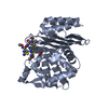 3hlwC 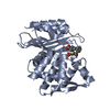 3hvfC  2p74S C: citing same article ( S: Starting model for refinement |
|---|---|
| Similar structure data |


-Links
 PDBj
PDBj


- Assembly
| Deposited unit | 
| ||||||||
|---|---|---|---|---|---|---|---|---|---|
| 1 | 
| ||||||||
| 2 | 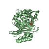
| ||||||||
| Unit cell |
|
-Components
| #1: Protein | Mass: 27942.467 Da / Num. of mol.: 2 / Fragment: UNP residues 29-291 / Mutation: S70G Source method: isolated from a genetically manipulated source Source: (gene. exp.) #2: Chemical |   Mass: 94.971 Da / Num. of mol.: 3 / Source method: obtained synthetically / Formula: PO4 Mass: 94.971 Da / Num. of mol.: 3 / Source method: obtained synthetically / Formula: PO4#3: Water | ChemComp-HOH / |  Mass: 18.015 Da / Num. of mol.: 932 / Source method: isolated from a natural source / Formula: H2O Mass: 18.015 Da / Num. of mol.: 932 / Source method: isolated from a natural source / Formula: H2OSequence details | RESIDUE NUMBERS 58, 239, 253 ARE SIMPLY SKIPPED. | |
|---|
-Experimental details
-Experiment
| Experiment | Method: X-RAY DIFFRACTION / Number of used crystals: 1 |
|---|
- Sample preparation
| Crystal | Density Matthews: 1.99 Å3/Da / Density % sol: 38.25 % |
|---|---|
| Crystal grow | Temperature: 293 K / Method: vapor diffusion, hanging drop / pH: 8.8 Details: potassium phosphate, pH 8.8, VAPOR DIFFUSION, HANGING DROP, temperature 293K |
-Data collection
| Diffraction | Mean temperature: 100 K |
|---|---|
| Diffraction source | Source: SYNCHROTRON / Site: ESRF  / Beamline: ID23-1 / Wavelength: 1.0723 Å / Beamline: ID23-1 / Wavelength: 1.0723 Å |
| Detector | Type: ADSC QUANTUM 315r / Detector: CCD / Date: Feb 8, 2008 / Details: Single Silicon (111) monochromator |
| Radiation | Monochromator: Single crystal / Protocol: SINGLE WAVELENGTH / Monochromatic (M) / Laue (L): M / Scattering type: x-ray |
| Radiation wavelength | Wavelength: 1.0723 Å / Relative weight: 1 |
| Reflection | Resolution: 1.4→50 Å / Num. all: 82496 / Num. obs: 78452 / % possible obs: 96.2 % / Observed criterion σ(I): 2 / Redundancy: 4.9 % / Biso Wilson estimate: 15.4 Å2 / Rmerge(I) obs: 0.047 / Net I/σ(I): 22.43 |
| Reflection shell | Resolution: 1.4→1.45 Å / Redundancy: 4.8 % / Rmerge(I) obs: 0.18 / Mean I/σ(I) obs: 5.43 / Num. unique all: 7779 / % possible all: 91 |
-Phasing
| Phasing | Method: molecular replacement |
|---|
- Processing
| Software |
| ||||||||||||||||||||||||||||||||||||||||||||||||||||||||||||||||||||||||||||||||||||||||||
|---|---|---|---|---|---|---|---|---|---|---|---|---|---|---|---|---|---|---|---|---|---|---|---|---|---|---|---|---|---|---|---|---|---|---|---|---|---|---|---|---|---|---|---|---|---|---|---|---|---|---|---|---|---|---|---|---|---|---|---|---|---|---|---|---|---|---|---|---|---|---|---|---|---|---|---|---|---|---|---|---|---|---|---|---|---|---|---|---|---|---|---|
| Refinement | Method to determine structure: MOLECULAR REPLACEMENT Starting model: PDB ENTRY 2P74 Resolution: 1.45→42.6 Å / Cor.coef. Fo:Fc: 0.961 / Cor.coef. Fo:Fc free: 0.946 / Occupancy max: 1 / Occupancy min: 0.3 / Cross valid method: THROUGHOUT / σ(F): 0 / ESU R: 0.072 / ESU R Free: 0.073 / Stereochemistry target values: MAXIMUM LIKELIHOOD
| ||||||||||||||||||||||||||||||||||||||||||||||||||||||||||||||||||||||||||||||||||||||||||
| Solvent computation | Ion probe radii: 0.8 Å / Shrinkage radii: 0.8 Å / VDW probe radii: 1.2 Å / Solvent model: MASK | ||||||||||||||||||||||||||||||||||||||||||||||||||||||||||||||||||||||||||||||||||||||||||
| Displacement parameters | Biso max: 298.1 Å2 / Biso mean: 10.506 Å2 / Biso min: 2 Å2
| ||||||||||||||||||||||||||||||||||||||||||||||||||||||||||||||||||||||||||||||||||||||||||
| Refine analyze | Luzzati coordinate error obs: 0.131 Å | ||||||||||||||||||||||||||||||||||||||||||||||||||||||||||||||||||||||||||||||||||||||||||
| Refinement step | Cycle: LAST / Resolution: 1.45→42.6 Å
| ||||||||||||||||||||||||||||||||||||||||||||||||||||||||||||||||||||||||||||||||||||||||||
| Refine LS restraints |
| ||||||||||||||||||||||||||||||||||||||||||||||||||||||||||||||||||||||||||||||||||||||||||
| LS refinement shell | Resolution: 1.45→1.488 Å / Total num. of bins used: 20
|
