 Movie
Movie Controller


+ Open data

- Basic information
| Entry | Database: PDB / ID: 6v2j | |||||||||
|---|---|---|---|---|---|---|---|---|---|---|
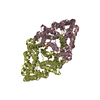
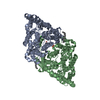
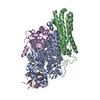
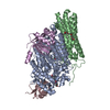
| Title | Crystal structure of ClC-ec1 triple mutant (E113Q, E148Q, E203Q) | |||||||||
 Components Components | H(+)/Cl(-) exchange transporter ClcA | |||||||||
 Keywords Keywords | TRANSPORT PROTEIN / chloride transporters / CLC-ec1 | |||||||||
| Function / homology |  Function and homology information Function and homology informationcellular stress response to acidic pH / chloride:proton antiporter activity / voltage-gated chloride channel activity / proton transmembrane transport / chloride transmembrane transport / identical protein binding / plasma membrane Similarity search - Function | |||||||||
| Biological species |  | |||||||||
| Method |  X-RAY DIFFRACTION / SYNCHROTRON / MOLECULAR REPLACEMENT / molecular replacement / Resolution: 2.62 Å X-RAY DIFFRACTION / SYNCHROTRON / MOLECULAR REPLACEMENT / molecular replacement / Resolution: 2.62 Å | |||||||||
 Authors Authors | Maduke, M. / Mathews, I.I. / Chavan, T.S. | |||||||||
| Funding support |  United States, 2items United States, 2items
| |||||||||
 Citation Citation | Journal: Elife / Year: 2020 Title: A CLC-ec1 mutant reveals global conformational change and suggests a unifying mechanism for the CLC Cl - /H + transport cycle. Authors: Chavan, T.S. / Cheng, R.C. / Jiang, T. / Mathews, I.I. / Stein, R.A. / Koehl, A. / Mchaourab, H.S. / Tajkhorshid, E. / Maduke, M. | |||||||||
| History |
|
- Structure visualization
| Structure viewer | Molecule: MolmilJmol/JSmol |
|---|
- Downloads & links
-Download
| PDBx/mmCIF format | 6v2j.cif.gz | 179.1 KB | Display | PDBx/mmCIF format |
|---|---|---|---|---|
| PDB format | pdb6v2j.ent.gz | 141.5 KB | Display | PDB format |
| PDBx/mmJSON format | 6v2j.json.gz | Tree view | PDBx/mmJSON format | |
| Others |  Other downloads Other downloads |
-Validation report
| Arichive directory | https://data.pdbj.org/pub/pdb/validation_reports/v2/6v2jftp://data.pdbj.org/pub/pdb/validation_reports/v2/6v2j | HTTPS FTP |
|---|
-Related structure data
| Related structure data | 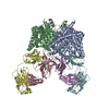 1otsS S: Starting model for refinement |
|---|---|
| Similar structure data |


-Links
 PDBj
PDBj- Assembly
| Deposited unit | 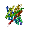
| ||||||||
|---|---|---|---|---|---|---|---|---|---|
| 1 | 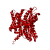
| ||||||||
| Unit cell |
|
-Components
| #1: Protein | Mass: 52112.402 Da / Num. of mol.: 1 / Mutation: E113Q, E148Q, E203Q Source method: isolated from a genetically manipulated source Source: (gene. exp.) Strain: K12 / Gene: clcA, eriC, yadQ, b0155, JW5012 / Production host: | ||||
|---|---|---|---|---|---|
| #2: Chemical |   Mass: 35.453 Da / Num. of mol.: 3 / Source method: obtained synthetically / Formula: Cl / Feature type: SUBJECT OF INVESTIGATION Mass: 35.453 Da / Num. of mol.: 3 / Source method: obtained synthetically / Formula: Cl / Feature type: SUBJECT OF INVESTIGATION#3: Water | ChemComp-HOH / |  Mass: 18.015 Da / Num. of mol.: 55 / Source method: isolated from a natural source / Formula: H2O Mass: 18.015 Da / Num. of mol.: 55 / Source method: isolated from a natural source / Formula: H2OHas ligand of interest | Y | |
-Experimental details
-Experiment
| Experiment | Method: X-RAY DIFFRACTION / Number of used crystals: 1 |
|---|
- Sample preparation
| Crystal | Density Matthews: 2.87 Å3/Da / Density % sol: 57.1 % |
|---|---|
| Crystal grow | Temperature: 289 K / Method: lipidic cubic phase / pH: 8.5 Details: 100mM Tris, 100mM sodium malonate, 30% PEG 400, 2.5% 2-Methyl-2,4-pentanediol |
-Data collection
| Diffraction | Mean temperature: 100 K / Serial crystal experiment: N |
|---|---|
| Diffraction source | Source: SYNCHROTRON / Site: APS / Beamline: 23-ID-D / Wavelength: 1.03321 Å |
| Detector | Type: DECTRIS PILATUS3 6M / Detector: PIXEL / Date: Nov 11, 2017 |
| Radiation | Monochromator: SI(111) / Protocol: SINGLE WAVELENGTH / Monochromatic (M) / Laue (L): M / Scattering type: x-ray |
| Radiation wavelength | Wavelength: 1.03321 Å / Relative weight: 1 |
| Reflection | Resolution: 2.62→28.65 Å / Num. obs: 17991 / % possible obs: 97.5 % / Redundancy: 7.4 % / CC1/2: 0.991 / Rmerge(I) obs: 0.173 / Rpim(I) all: 0.086 / Rrim(I) all: 0.185 / Net I/σ(I): 8.4 |
| Reflection shell | Resolution: 2.62→2.73 Å / Rmerge(I) obs: 0.866 / Num. unique obs: 1830 / CC1/2: 0.831 / Rrim(I) all: 1 |
-Phasing
| Phasing | Method: molecular replacement |
|---|
- Processing
| Software |
| |||||||||||||||||||||||||||||||||||||||||||||||||||||||||||||||||
|---|---|---|---|---|---|---|---|---|---|---|---|---|---|---|---|---|---|---|---|---|---|---|---|---|---|---|---|---|---|---|---|---|---|---|---|---|---|---|---|---|---|---|---|---|---|---|---|---|---|---|---|---|---|---|---|---|---|---|---|---|---|---|---|---|---|---|
| Refinement | Method to determine structure: MOLECULAR REPLACEMENT Starting model: 1OTS Resolution: 2.62→28.65 Å / Cor.coef. Fo:Fc: 0.952 / Cor.coef. Fo:Fc free: 0.916 / WRfactor Rfree: 0.2534 / WRfactor Rwork: 0.1844 / FOM work R set: 0.756 / SU B: 31.132 / SU ML: 0.294 / SU R Cruickshank DPI: 0.2717 / SU Rfree: 0.3105 / Cross valid method: THROUGHOUT / σ(F): 0 / ESU R Free: 0.311 / Stereochemistry target values: MAXIMUM LIKELIHOOD Details: HYDROGENS HAVE BEEN ADDED IN THE RIDING POSITIONS U VALUES : WITH TLS ADDED
| |||||||||||||||||||||||||||||||||||||||||||||||||||||||||||||||||
| Solvent computation | Ion probe radii: 0.8 Å / Shrinkage radii: 0.8 Å / VDW probe radii: 1.2 Å / Solvent model: MASK | |||||||||||||||||||||||||||||||||||||||||||||||||||||||||||||||||
| Displacement parameters | Biso max: 144.27 Å2 / Biso mean: 60.26 Å2 / Biso min: 41.9 Å2
| |||||||||||||||||||||||||||||||||||||||||||||||||||||||||||||||||
| Refinement step | Cycle: final / Resolution: 2.62→28.65 Å
| |||||||||||||||||||||||||||||||||||||||||||||||||||||||||||||||||
| Refine LS restraints |
| |||||||||||||||||||||||||||||||||||||||||||||||||||||||||||||||||
| LS refinement shell | Resolution: 2.62→2.683 Å / Rfactor Rfree error: 0
| |||||||||||||||||||||||||||||||||||||||||||||||||||||||||||||||||
| Refinement TLS params. | Method: refined / Origin x: 35.5522 Å / Origin y: 28.1049 Å / Origin z: -18.3937 Å
| |||||||||||||||||||||||||||||||||||||||||||||||||||||||||||||||||
| Refinement TLS group |
|
