 Movie
Movie Controller

[English] 日本語
 Yorodumi
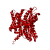
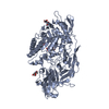
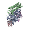
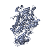
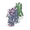
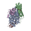
- PDB-1kpl: Crystal Structure of the ClC Chloride Channel from S. typhimurium -
+ Open data

- Basic information
| Entry | Database: PDB / ID: 1kpl | ||||||
|---|---|---|---|---|---|---|---|
| Title | Crystal Structure of the ClC Chloride Channel from S. typhimurium | ||||||
 Components Components | putative ClC family, chlorine transport protein | ||||||
 Keywords Keywords | MEMBRANE PROTEIN / helical membrane protein / homodimer / ion channel | ||||||
| Function / homology |  Function and homology information Function and homology informationvoltage-gated chloride channel activity / antiporter activity / plasma membrane Similarity search - Function | ||||||
| Biological species |  Salmonella typhimurium (bacteria) Salmonella typhimurium (bacteria) | ||||||
| Method |  X-RAY DIFFRACTION / SYNCHROTRON / SIRAS / Resolution: 3 Å X-RAY DIFFRACTION / SYNCHROTRON / SIRAS / Resolution: 3 Å | ||||||
 Authors Authors | Dutzler, R. / Campbell, E.B. / Cadene, M. / Chait, B.T. / MacKinnon, R. | ||||||
 Citation Citation | Journal: Nature / Year: 2002 Title: X-ray structure of a ClC chloride channel at 3.0 A reveals the molecular basis of anion selectivity. Authors: Dutzler, R. / Campbell, E.B. / Cadene, M. / Chait, B.T. / MacKinnon, R. | ||||||
| History |
|
- Structure visualization
| Structure viewer | Molecule: MolmilJmol/JSmol |
|---|
- Downloads & links
-Download
| PDBx/mmCIF format | 1kpl.cif.gz | 326.8 KB | Display | PDBx/mmCIF format |
|---|---|---|---|---|
| PDB format | pdb1kpl.ent.gz | 269.4 KB | Display | PDB format |
| PDBx/mmJSON format | 1kpl.json.gz | Tree view | PDBx/mmJSON format | |
| Others |  Other downloads Other downloads |
-Validation report
| Arichive directory | https://data.pdbj.org/pub/pdb/validation_reports/kp/1kplftp://data.pdbj.org/pub/pdb/validation_reports/kp/1kpl | HTTPS FTP |
|---|
-Related structure data


-Links
 PDBj
PDBj

- Assembly
| Deposited unit | 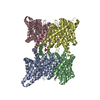
| ||||||||||||
|---|---|---|---|---|---|---|---|---|---|---|---|---|---|
| 1 | 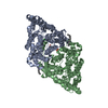
| ||||||||||||
| 2 | 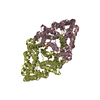
| ||||||||||||
| Unit cell |
| ||||||||||||
| Noncrystallographic symmetry (NCS) | NCS oper:
| ||||||||||||
| Details | The biologically active assembly is a homodimer. The two biological dimers in the asymmetric unit are composed of protein chains A and B, C and D. Strict crystallograhic NCS constraints have been maintained between the two dimers in the asymmetric unit. |
-Components
-Protein , 1 types, 4 molecules ABCD
| #1: Protein | Mass: 50499.586 Da / Num. of mol.: 4 / Mutation: M26L/C264V Source method: isolated from a genetically manipulated source Source: (gene. exp.) Salmonella typhimurium (bacteria) / Plasmid: pET28b+ / Species (production host): Escherichia coli / Production host: |
|---|
-Non-polymers , 5 types, 14 molecules 


| #2: Chemical | ChemComp-CL /  Mass: 35.453 Da / Num. of mol.: 4 / Source method: obtained synthetically / Formula: Cl Mass: 35.453 Da / Num. of mol.: 4 / Source method: obtained synthetically / Formula: Cl#3: Chemical | ChemComp-SO4 /  Mass: 96.063 Da / Num. of mol.: 4 / Source method: obtained synthetically / Formula: SO4 Mass: 96.063 Da / Num. of mol.: 4 / Source method: obtained synthetically / Formula: SO4#4: Chemical |  Mass: 114.229 Da / Num. of mol.: 2 / Source method: obtained synthetically / Formula: C8H18 Mass: 114.229 Da / Num. of mol.: 2 / Source method: obtained synthetically / Formula: C8H18#5: Chemical |  Mass: 212.415 Da / Num. of mol.: 2 / Source method: obtained synthetically / Formula: C15H32 Mass: 212.415 Da / Num. of mol.: 2 / Source method: obtained synthetically / Formula: C15H32#6: Water | ChemComp-HOH / | Mass: 18.015 Da / Num. of mol.: 2 / Source method: isolated from a natural source / Formula: H2O |
|---|
-Experimental details
-Experiment
| Experiment | Method: X-RAY DIFFRACTION |
|---|
- Sample preparation
| Crystal | Density Matthews: 3.3 Å3/Da / Density % sol: 62.78 % | |||||||||||||||||||||||||||||||||||||||||||||||||||||||||||||||
|---|---|---|---|---|---|---|---|---|---|---|---|---|---|---|---|---|---|---|---|---|---|---|---|---|---|---|---|---|---|---|---|---|---|---|---|---|---|---|---|---|---|---|---|---|---|---|---|---|---|---|---|---|---|---|---|---|---|---|---|---|---|---|---|---|
| Crystal grow | Temperature: 293 K / Method: vapor diffusion, sitting drop / pH: 4.6 Details: PEG 400, acetate, sodium sulfate, lithium sulfate, pH 4.6, VAPOR DIFFUSION, SITTING DROP, temperature 293K | |||||||||||||||||||||||||||||||||||||||||||||||||||||||||||||||
| Crystal grow | *PLUS Temperature: 20 ℃ / pH: 7.5 | |||||||||||||||||||||||||||||||||||||||||||||||||||||||||||||||
| Components of the solutions | *PLUS
|
-Data collection
| Diffraction | Mean temperature: 100 K |
|---|---|
| Diffraction source | Source: SYNCHROTRON / Site: NSLS  / Beamline: X25 / Wavelength: 1.1 Å / Beamline: X25 / Wavelength: 1.1 Å |
| Detector | Type: BRANDEIS / Detector: CCD / Date: Oct 3, 2001 |
| Radiation | Protocol: SINGLE WAVELENGTH / Monochromatic (M) / Laue (L): M / Scattering type: x-ray |
| Radiation wavelength | Wavelength: 1.1 Å / Relative weight: 1 |
| Reflection | Resolution: 3→35 Å / Num. all: 53397 / Num. obs: 50567 / % possible obs: 94.7 % / Observed criterion σ(F): 1 / Observed criterion σ(I): 1 / Redundancy: 2.4 % / Biso Wilson estimate: 85.1 Å2 / Rmerge(I) obs: 0.065 / Net I/σ(I): 13.7 |
| Reflection shell | Resolution: 3→3.11 Å / Rmerge(I) obs: 0.544 / Mean I/σ(I) obs: 1.7 / % possible all: 96.6 |
| Reflection shell | *PLUS % possible obs: 96.6 % |
- Processing
| Software |
| ||||||||||||||||||||||||||||||||||||||||||||
|---|---|---|---|---|---|---|---|---|---|---|---|---|---|---|---|---|---|---|---|---|---|---|---|---|---|---|---|---|---|---|---|---|---|---|---|---|---|---|---|---|---|---|---|---|---|
| Refinement | Method to determine structure: SIRAS / Resolution: 3→20 Å / Rfactor Rfree error: 0.004 / Data cutoff high absF: 1592586.14 / Data cutoff low absF: 0 / Isotropic thermal model: RESTRAINED / Cross valid method: THROUGHOUT / σ(F): 0 / Stereochemistry target values: Engh & Huber Details: Strict 2-fold NCS constraints between the two dimers in the asymmetric unit were maintained. BULK SOLVENT MODEL USED.
| ||||||||||||||||||||||||||||||||||||||||||||
| Displacement parameters | Biso mean: 79.6 Å2
| ||||||||||||||||||||||||||||||||||||||||||||
| Refine analyze |
| ||||||||||||||||||||||||||||||||||||||||||||
| Refinement step | Cycle: LAST / Resolution: 3→20 Å
| ||||||||||||||||||||||||||||||||||||||||||||
| Refine LS restraints |
| ||||||||||||||||||||||||||||||||||||||||||||
| Refine LS restraints NCS | NCS model details: CONSTR | ||||||||||||||||||||||||||||||||||||||||||||
| LS refinement shell | Resolution: 3→3.19 Å / Rfactor Rfree error: 0.014 / Total num. of bins used: 6
| ||||||||||||||||||||||||||||||||||||||||||||
| Xplor file |
| ||||||||||||||||||||||||||||||||||||||||||||
| Software | *PLUS Name: CNS / Version: 0.3 / Classification: refinement | ||||||||||||||||||||||||||||||||||||||||||||
| Refinement | *PLUS σ(F): 0 / % reflection Rfree: 10 % / Rfactor Rfree: 0.288 | ||||||||||||||||||||||||||||||||||||||||||||
| Solvent computation | *PLUS | ||||||||||||||||||||||||||||||||||||||||||||
| Displacement parameters | *PLUS Biso mean: 79.6 Å2 | ||||||||||||||||||||||||||||||||||||||||||||
| Refine LS restraints | *PLUS
| ||||||||||||||||||||||||||||||||||||||||||||
| LS refinement shell | *PLUS Rfactor Rfree: 0.386 / % reflection Rfree: 9.7 % / Rfactor Rwork: 0.361 |
