 Movie
Movie Controller

[English] 日本語
 Yorodumi
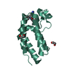
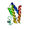
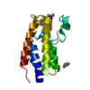
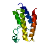
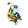
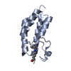
- PDB-6v0x: Crystal structure of the bromodomain of human BRD9 bound to sunitinib -
+ Open data

- Basic information
| Entry | Database: PDB / ID: 6v0x | ||||||
|---|---|---|---|---|---|---|---|
| Title | Crystal structure of the bromodomain of human BRD9 bound to sunitinib | ||||||
 Components Components | Bromodomain-containing protein 9 | ||||||
 Keywords Keywords | GENE REGULATION / Sunitinib / BRD9 / mSWI/SNF / BAF / non-BET / BRD | ||||||
| Function / homology |  Function and homology information Function and homology informationFormation of the non-canonical BAF (ncBAF) complex / GBAF complex / SWI/SNF complex / positive regulation of stem cell population maintenance / negative regulation of cell differentiation / histone reader activity / double-strand break repair via homologous recombination / site of double-strand break / nucleic acid binding / protein-macromolecule adaptor activity ...Formation of the non-canonical BAF (ncBAF) complex / GBAF complex / SWI/SNF complex / positive regulation of stem cell population maintenance / negative regulation of cell differentiation / histone reader activity / double-strand break repair via homologous recombination / site of double-strand break / nucleic acid binding / protein-macromolecule adaptor activity / chromatin remodeling / positive regulation of cell population proliferation / regulation of transcription by RNA polymerase II / chromatin / nucleoplasm / nucleus Similarity search - Function | ||||||
| Biological species |  Homo sapiens (human) Homo sapiens (human) | ||||||
| Method |  X-RAY DIFFRACTION / SYNCHROTRON / MOLECULAR REPLACEMENT / Resolution: 1.5 Å X-RAY DIFFRACTION / SYNCHROTRON / MOLECULAR REPLACEMENT / Resolution: 1.5 Å | ||||||
 Authors Authors | Karim, M.R. / Chan, A. / Zhu, J.Y. / Schonbrunn, E. | ||||||
 Citation Citation | Journal: J.Med.Chem. / Year: 2020 Title: Structural Basis of Inhibitor Selectivity in the BRD7/9 Subfamily of Bromodomains. Authors: Karim, R.M. / Chan, A. / Zhu, J.Y. / Schonbrunn, E. | ||||||
| History |
|
- Structure visualization
| Structure viewer | Molecule: MolmilJmol/JSmol |
|---|
- Downloads & links
-Download
| PDBx/mmCIF format | 6v0x.cif.gz | 60.3 KB | Display | PDBx/mmCIF format |
|---|---|---|---|---|
| PDB format | pdb6v0x.ent.gz | 41.6 KB | Display | PDB format |
| PDBx/mmJSON format | 6v0x.json.gz | Tree view | PDBx/mmJSON format | |
| Others |  Other downloads Other downloads |
-Validation report
| Arichive directory | https://data.pdbj.org/pub/pdb/validation_reports/v0/6v0xftp://data.pdbj.org/pub/pdb/validation_reports/v0/6v0x | HTTPS FTP |
|---|
-Related structure data
| Related structure data |  6ppaC 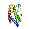 6uzfSC  6v0qC  6v0sC  6v0uC 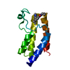 6v14C  6v16C  6v17C  6v1bC  6v1eC  6v1fC  6v1hC  6v1kC  6v1lC  6v1uC S: Starting model for refinement C: citing same article ( |
|---|---|
| Similar structure data |

-Links
 PDBj
PDBj- Assembly
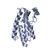
| Deposited unit | 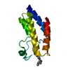
| ||||||||
|---|---|---|---|---|---|---|---|---|---|
| 1 |
| ||||||||
| Unit cell |
|
-Components
| #1: Protein | Mass: 14249.763 Da / Num. of mol.: 1 Source method: isolated from a genetically manipulated source Source: (gene. exp.) Homo sapiens (human) / Gene: BRD9, UNQ3040/PRO9856 / Plasmid: pNIC28-Bsa4 / Production host:  |
|---|---|
| #2: Chemical | ChemComp-B49 / 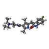  Mass: 398.474 Da / Num. of mol.: 1 / Source method: obtained synthetically / Formula: C22H27FN4O2 / Feature type: SUBJECT OF INVESTIGATION Mass: 398.474 Da / Num. of mol.: 1 / Source method: obtained synthetically / Formula: C22H27FN4O2 / Feature type: SUBJECT OF INVESTIGATION |
| #3: Water | ChemComp-HOH /  Mass: 18.015 Da / Num. of mol.: 91 / Source method: isolated from a natural source / Formula: H2O Mass: 18.015 Da / Num. of mol.: 91 / Source method: isolated from a natural source / Formula: H2O |
| Has ligand of interest | Y |
-Experimental details
-Experiment
| Experiment | Method: X-RAY DIFFRACTION / Number of used crystals: 1 |
|---|
- Sample preparation
| Crystal | Density Matthews: 2.37 Å3/Da / Density % sol: 48.08 % |
|---|---|
| Crystal grow | Temperature: 291 K / Method: vapor diffusion, sitting drop Details: 0.2 M NaCl, 0.1 M Bis-Tris (pH 5.5), and 25% PEG 3350 |
-Data collection
| Diffraction | Mean temperature: 100 K / Serial crystal experiment: N | ||||||||||||||||||||||||||||||
|---|---|---|---|---|---|---|---|---|---|---|---|---|---|---|---|---|---|---|---|---|---|---|---|---|---|---|---|---|---|---|---|
| Diffraction source | Source: SYNCHROTRON / Site: APS  / Beamline: 22-ID / Wavelength: 1 Å / Beamline: 22-ID / Wavelength: 1 Å | ||||||||||||||||||||||||||||||
| Detector | Type: DECTRIS EIGER X 16M / Detector: PIXEL / Date: Oct 27, 2018 | ||||||||||||||||||||||||||||||
| Radiation | Monochromator: Si filter / Protocol: SINGLE WAVELENGTH / Monochromatic (M) / Laue (L): M / Scattering type: x-ray | ||||||||||||||||||||||||||||||
| Radiation wavelength | Wavelength: 1 Å / Relative weight: 1 | ||||||||||||||||||||||||||||||
| Reflection | Resolution: 1.5→32.53 Å / Num. obs: 18344 / % possible obs: 97.9 % / Redundancy: 13.1 % / CC1/2: 0.994 / Rmerge(I) obs: 0.109 / Rpim(I) all: 0.032 / Rrim(I) all: 0.114 / Net I/σ(I): 22.6 / Num. measured all: 239696 / Scaling rejects: 350 | ||||||||||||||||||||||||||||||
| Reflection shell | Diffraction-ID: 1
|
- Processing
| Software |
| |||||||||||||||||||||||||||||||||||||||||||||||||||||||||||||||||||||||||||||||||||||||||||||||||||||||||||||||||||||||||||||
|---|---|---|---|---|---|---|---|---|---|---|---|---|---|---|---|---|---|---|---|---|---|---|---|---|---|---|---|---|---|---|---|---|---|---|---|---|---|---|---|---|---|---|---|---|---|---|---|---|---|---|---|---|---|---|---|---|---|---|---|---|---|---|---|---|---|---|---|---|---|---|---|---|---|---|---|---|---|---|---|---|---|---|---|---|---|---|---|---|---|---|---|---|---|---|---|---|---|---|---|---|---|---|---|---|---|---|---|---|---|---|---|---|---|---|---|---|---|---|---|---|---|---|---|---|---|---|
| Refinement | Method to determine structure: MOLECULAR REPLACEMENT Starting model: 6UZF Resolution: 1.5→31.241 Å / SU ML: 0.13 / Cross valid method: THROUGHOUT / σ(F): 1.35 / Phase error: 24.16 / Stereochemistry target values: ML
| |||||||||||||||||||||||||||||||||||||||||||||||||||||||||||||||||||||||||||||||||||||||||||||||||||||||||||||||||||||||||||||
| Solvent computation | Shrinkage radii: 0.9 Å / VDW probe radii: 1.11 Å / Solvent model: FLAT BULK SOLVENT MODEL | |||||||||||||||||||||||||||||||||||||||||||||||||||||||||||||||||||||||||||||||||||||||||||||||||||||||||||||||||||||||||||||
| Displacement parameters | Biso max: 100.99 Å2 / Biso mean: 38.164 Å2 / Biso min: 18.75 Å2 | |||||||||||||||||||||||||||||||||||||||||||||||||||||||||||||||||||||||||||||||||||||||||||||||||||||||||||||||||||||||||||||
| Refinement step | Cycle: final / Resolution: 1.5→31.241 Å
| |||||||||||||||||||||||||||||||||||||||||||||||||||||||||||||||||||||||||||||||||||||||||||||||||||||||||||||||||||||||||||||
| LS refinement shell | Refine-ID: X-RAY DIFFRACTION / Rfactor Rfree error: 0
| |||||||||||||||||||||||||||||||||||||||||||||||||||||||||||||||||||||||||||||||||||||||||||||||||||||||||||||||||||||||||||||
| Refinement TLS params. | Method: refined / Refine-ID: X-RAY DIFFRACTION
| |||||||||||||||||||||||||||||||||||||||||||||||||||||||||||||||||||||||||||||||||||||||||||||||||||||||||||||||||||||||||||||
| Refinement TLS group |
|
