 Movie
Movie Controller

[English] 日本語
 Yorodumi
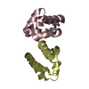
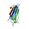
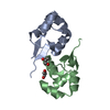
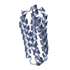
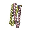
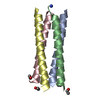
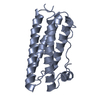
- PDB-6uvi: Crystal structure of alr1298, a pentapeptide repeat protein from ... -
+ Open data

- Basic information
| Entry | Database: PDB / ID: 6uvi | ||||||
|---|---|---|---|---|---|---|---|
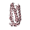
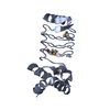
| Title | Crystal structure of alr1298, a pentapeptide repeat protein from Nostoc Pcc 7120, determined at 2.3 Angstrom resolution | ||||||
 Components Components | Alr1298 protein | ||||||
 Keywords Keywords | UNKNOWN FUNCTION / pentapeptide repeat / protein repeat / five residue right-handed-beta helix | ||||||
| Function / homology | : / Pentapeptide repeats (8 copies) / Pentapeptide repeat / Alr1298 protein Function and homology information Function and homology information | ||||||
| Biological species |  Nostoc sp. PCC 7120 (bacteria) Nostoc sp. PCC 7120 (bacteria) | ||||||
| Method |  X-RAY DIFFRACTION / SYNCHROTRON / MOLECULAR REPLACEMENT / Resolution: 2.1 Å X-RAY DIFFRACTION / SYNCHROTRON / MOLECULAR REPLACEMENT / Resolution: 2.1 Å | ||||||
 Authors Authors | Kennedy, M.A. / Zhang, R. | ||||||
 Citation Citation | Journal: Proteins / Year: 2020 Title: Crystal structure of Alr1298, a pentapeptide repeat protein from the cyanobacterium Nostoc sp. PCC 7120, determined at 2.1 angstrom resolution. Authors: Zhang, R. / Ni, S. / Kennedy, M.A. | ||||||
| History |
|
- Structure visualization
| Structure viewer | Molecule: MolmilJmol/JSmol |
|---|
- Downloads & links
-Download
| PDBx/mmCIF format | 6uvi.cif.gz | 151.3 KB | Display | PDBx/mmCIF format |
|---|---|---|---|---|
| PDB format | pdb6uvi.ent.gz | 98.2 KB | Display | PDB format |
| PDBx/mmJSON format | 6uvi.json.gz | Tree view | PDBx/mmJSON format | |
| Others |  Other downloads Other downloads |
-Validation report
| Arichive directory | https://data.pdbj.org/pub/pdb/validation_reports/uv/6uviftp://data.pdbj.org/pub/pdb/validation_reports/uv/6uvi | HTTPS FTP |
|---|
-Related structure data
| Related structure data | 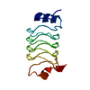 6omxC  6uv7SC S: Starting model for refinement C: citing same article ( |
|---|---|
| Similar structure data |
-Links
 PDBj
PDBj- Assembly
| Deposited unit | 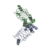
| ||||||||||||
|---|---|---|---|---|---|---|---|---|---|---|---|---|---|
| 1 | 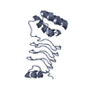
| ||||||||||||
| 2 | 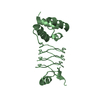
| ||||||||||||
| Unit cell |
|
-Components
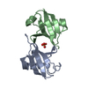
| #1: Protein | Mass: 20954.623 Da / Num. of mol.: 2 Source method: isolated from a genetically manipulated source Source: (gene. exp.) Nostoc sp. PCC 7120 (bacteria) / Strain: PCC 7120 / SAG 25.82 / UTEX 2576 / Gene: alr1298 / Production host: #2: Water | ChemComp-HOH / |  Mass: 18.015 Da / Num. of mol.: 68 / Source method: isolated from a natural source / Formula: H2O Mass: 18.015 Da / Num. of mol.: 68 / Source method: isolated from a natural source / Formula: H2O |
|---|
-Experimental details
-Experiment
| Experiment | Method: X-RAY DIFFRACTION / Number of used crystals: 1 |
|---|
- Sample preparation
| Crystal | Density Matthews: 2.24 Å3/Da / Density % sol: 45.2 % |
|---|---|
| Crystal grow | Temperature: 298 K / Method: vapor diffusion, hanging drop Details: 0.2 M Ammonium acetate, 0.1 M Sodium acetate trihydrate pH 4.6, 30% w/v Polyethylene glycol 4,000 |
-Data collection
| Diffraction | Mean temperature: 100 K / Serial crystal experiment: N |
|---|---|
| Diffraction source | Source: SYNCHROTRON / Site: APS  / Beamline: 31-ID / Wavelength: 0.97931 Å / Beamline: 31-ID / Wavelength: 0.97931 Å |
| Detector | Type: DECTRIS PILATUS3 S 6M / Detector: PIXEL / Date: Dec 6, 2018 |
| Radiation | Protocol: SINGLE WAVELENGTH / Monochromatic (M) / Laue (L): M / Scattering type: x-ray |
| Radiation wavelength | Wavelength: 0.97931 Å / Relative weight: 1 |
| Reflection | Resolution: 2.1→31.32 Å / Num. obs: 22287 / % possible obs: 98.03 % / Redundancy: 2 % / Biso Wilson estimate: 40.41 Å2 / CC1/2: 1 / Rmerge(I) obs: 0.01859 / Rpim(I) all: 0.01859 / Rrim(I) all: 0.02629 / Net I/σ(I): 18.53 |
| Reflection shell | Resolution: 2.1→2.21 Å / Rmerge(I) obs: 0.065 / Mean I/σ(I) obs: 17.7 / Num. unique obs: 22336 / Rpim(I) all: 0.028 / Rrim(I) all: 0.071 |
- Processing
| Software |
| |||||||||||||||||||||||||||||||||||||||||||||||||||||||||||||||||||||||||||||||||||||||||||||||||||||||||
|---|---|---|---|---|---|---|---|---|---|---|---|---|---|---|---|---|---|---|---|---|---|---|---|---|---|---|---|---|---|---|---|---|---|---|---|---|---|---|---|---|---|---|---|---|---|---|---|---|---|---|---|---|---|---|---|---|---|---|---|---|---|---|---|---|---|---|---|---|---|---|---|---|---|---|---|---|---|---|---|---|---|---|---|---|---|---|---|---|---|---|---|---|---|---|---|---|---|---|---|---|---|---|---|---|---|---|
| Refinement | Method to determine structure: MOLECULAR REPLACEMENT Starting model: 6UV7 Resolution: 2.1→31.32 Å / SU ML: 0.3238 / Cross valid method: FREE R-VALUE / σ(F): 1.34 / Phase error: 32.8294
| |||||||||||||||||||||||||||||||||||||||||||||||||||||||||||||||||||||||||||||||||||||||||||||||||||||||||
| Solvent computation | Shrinkage radii: 0.9 Å / VDW probe radii: 1.11 Å | |||||||||||||||||||||||||||||||||||||||||||||||||||||||||||||||||||||||||||||||||||||||||||||||||||||||||
| Displacement parameters | Biso mean: 60.16 Å2 | |||||||||||||||||||||||||||||||||||||||||||||||||||||||||||||||||||||||||||||||||||||||||||||||||||||||||
| Refinement step | Cycle: LAST / Resolution: 2.1→31.32 Å
| |||||||||||||||||||||||||||||||||||||||||||||||||||||||||||||||||||||||||||||||||||||||||||||||||||||||||
| Refine LS restraints |
| |||||||||||||||||||||||||||||||||||||||||||||||||||||||||||||||||||||||||||||||||||||||||||||||||||||||||
| LS refinement shell |
|
