 Movie
Movie Controller


+ Open data

- Basic information
| Entry | Database: PDB / ID: 1skv | ||||||
|---|---|---|---|---|---|---|---|
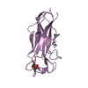
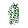
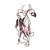
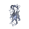
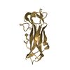
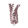
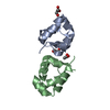
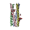
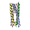
| Title | Crystal Structure of D-63 from Sulfolobus Spindle Virus 1 | ||||||
 Components Components | Hypothetical 7.5 kDa protein | ||||||
 Keywords Keywords | VIRAL PROTEIN / Sulfolobus Spindle Virus / SSV / Archaeal / Crenarchaeal / Helix-Turn-Helix / Four Helix Bundle | ||||||
| Function / homology | Hypothetical protein D-63 / : / Sulfolobus spindle-shape virus 1, protein D-63 / Helix hairpin bin / Helix hairpin bin domain superfamily / Helix Hairpins / Orthogonal Bundle / Mainly Alpha / Protein D-63 Function and homology information Function and homology information | ||||||
| Biological species |   Sulfolobus virus 1 Sulfolobus virus 1 | ||||||
| Method |  X-RAY DIFFRACTION / SYNCHROTRON / MAD / Resolution: 2.6 Å X-RAY DIFFRACTION / SYNCHROTRON / MAD / Resolution: 2.6 Å | ||||||
 Authors Authors | Kraft, P. / Kummel, D. / Oeckinghaus, A. / Gauss, G.H. / Wiedenheft, B. / Young, M. / Lawrence, C.M. | ||||||
 Citation Citation | Journal: J.Virol. / Year: 2004 Title: Structure of d-63 from sulfolobus spindle-shaped virus 1: surface properties of the dimeric four-helix bundle suggest an adaptor protein function Authors: Kraft, P. / Oeckinghaus, A. / Gauss, G.H. / Wiedenheft, B. / Young, M. / Lawrence, C.M. | ||||||
| History |
|
- Structure visualization
| Structure viewer | Molecule: MolmilJmol/JSmol |
|---|
- Downloads & links
-Download
| PDBx/mmCIF format | 1skv.cif.gz | 60 KB | Display | PDBx/mmCIF format |
|---|---|---|---|---|
| PDB format | pdb1skv.ent.gz | 46.1 KB | Display | PDB format |
| PDBx/mmJSON format | 1skv.json.gz | Tree view | PDBx/mmJSON format | |
| Others |  Other downloads Other downloads |
-Validation report
| Arichive directory | https://data.pdbj.org/pub/pdb/validation_reports/sk/1skvftp://data.pdbj.org/pub/pdb/validation_reports/sk/1skv | HTTPS FTP |
|---|
-Related structure data
| Similar structure data |
|---|

-Links
 PDBj
PDBj- Assembly
| Deposited unit | 
| ||||||||
|---|---|---|---|---|---|---|---|---|---|
| 1 | 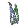
| ||||||||
| 2 | 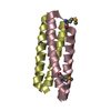
| ||||||||
| Unit cell |
| ||||||||
| Details | The biological assembly is the AB dimer / The biological assembly is the CD dimer |
-Components
| #1: Protein | Mass: 8402.422 Da / Num. of mol.: 4 Source method: isolated from a genetically manipulated source Source: (gene. exp.) Sulfolobus virus 1 / Genus: Fusellovirus / Gene: D-63 / Plasmid: pDest14 / Production host:  #2: Water | ChemComp-HOH / |  Mass: 18.015 Da / Num. of mol.: 10 / Source method: isolated from a natural source / Formula: H2O Mass: 18.015 Da / Num. of mol.: 10 / Source method: isolated from a natural source / Formula: H2OHas protein modification | Y | |
|---|
-Experimental details
-Experiment
| Experiment | Method: X-RAY DIFFRACTION / Number of used crystals: 1 |
|---|
- Sample preparation
| Crystal | Density Matthews: 2.99 Å3/Da / Density % sol: 58.48 % |
|---|---|
| Crystal grow | Temperature: 291 K / Method: vapor diffusion, hanging drop / pH: 5 Details: Ammonium Sulfate, ammonium acetate, PEG 4000, NaCl, Tris, pH 5.0, VAPOR DIFFUSION, HANGING DROP, temperature 291K |
-Data collection
| Diffraction | Mean temperature: 100 K | ||||||||||||
|---|---|---|---|---|---|---|---|---|---|---|---|---|---|
| Diffraction source | Source: SYNCHROTRON / Site: APS  / Beamline: 14-BM-D / Wavelength: 0.9778, 0.9787, 0.9789 / Beamline: 14-BM-D / Wavelength: 0.9778, 0.9787, 0.9789 | ||||||||||||
| Detector | Type: ADSC QUANTUM 4 / Detector: CCD / Date: Aug 16, 2002 | ||||||||||||
| Radiation | Protocol: MAD / Monochromatic (M) / Laue (L): M / Scattering type: x-ray | ||||||||||||
| Radiation wavelength |
| ||||||||||||
| Reflection | Resolution: 2.6→20 Å / Num. all: 10225 / Num. obs: 10095 / % possible obs: 98.7 % / Observed criterion σ(F): -1.5 / Observed criterion σ(I): -3 / Redundancy: 8.6 % / Rmerge(I) obs: 0.043 / Net I/σ(I): 42 | ||||||||||||
| Reflection shell | Resolution: 2.6→2.7 Å / Redundancy: 9.7 % / Rmerge(I) obs: 0.212 / Mean I/σ(I) obs: 9 / Num. unique all: 1121 / % possible all: 99.5 |
- Processing
| Software |
| |||||||||||||||||||||||||
|---|---|---|---|---|---|---|---|---|---|---|---|---|---|---|---|---|---|---|---|---|---|---|---|---|---|---|
| Refinement | Method to determine structure: MAD / Resolution: 2.6→15 Å / Isotropic thermal model: isotropic / Cross valid method: THROUGHOUT / σ(F): 0 / σ(I): 0 / Stereochemistry target values: Engh & Huber
| |||||||||||||||||||||||||
| Displacement parameters | Biso mean: 58 Å2
| |||||||||||||||||||||||||
| Refine analyze |
| |||||||||||||||||||||||||
| Refinement step | Cycle: LAST / Resolution: 2.6→15 Å
| |||||||||||||||||||||||||
| Refine LS restraints |
|
