 Movie
Movie Controller

[English] 日本語
 Yorodumi
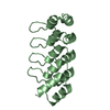
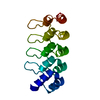
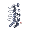
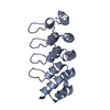
- PDB-6zz1: Crystal structure of MLKL executioner domain in complex with a co... -
+ Open data

- Basic information
| Entry | Database: PDB / ID: 6zz1 | ||||||
|---|---|---|---|---|---|---|---|
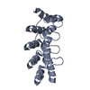
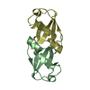
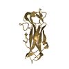
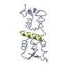
| Title | Crystal structure of MLKL executioner domain in complex with a covalent inhibitor | ||||||
 Components Components | Mixed lineage kinase domain-like protein | ||||||
 Keywords Keywords | LIPID BINDING PROTEIN / Necroptosis | ||||||
| Function / homology |  Function and homology information Function and homology informationexecution phase of necroptosis / Microbial modulation of RIPK1-mediated regulated necrosis / necroptotic signaling pathway / RIPK1-mediated regulated necrosis / necroptotic process / protein homotrimerization / TRP channels / Regulation of necroptotic cell death / cell junction / defense response to virus ...execution phase of necroptosis / Microbial modulation of RIPK1-mediated regulated necrosis / necroptotic signaling pathway / RIPK1-mediated regulated necrosis / necroptotic process / protein homotrimerization / TRP channels / Regulation of necroptotic cell death / cell junction / defense response to virus / cell surface receptor signaling pathway / protein kinase binding / protein-containing complex binding / ATP binding / identical protein binding / nucleus / plasma membrane / cytosol / cytoplasm Similarity search - Function | ||||||
| Biological species |  Homo sapiens (human) Homo sapiens (human) | ||||||
| Method |  X-RAY DIFFRACTION / SYNCHROTRON / MOLECULAR REPLACEMENT / Resolution: 1.64 Å X-RAY DIFFRACTION / SYNCHROTRON / MOLECULAR REPLACEMENT / Resolution: 1.64 Å | ||||||
 Authors Authors | Fiegen, D. / Bauer, M. / Nar, H. | ||||||
 Citation Citation | Journal: Proc.Natl.Acad.Sci.USA / Year: 2020 Title: Locking mixed-lineage kinase domain-like protein in its auto-inhibited state prevents necroptosis. Authors: Rubbelke, M. / Fiegen, D. / Bauer, M. / Binder, F. / Hamilton, J. / King, J. / Thamm, S. / Nar, H. / Zeeb, M. | ||||||
| History |
|
- Structure visualization
| Structure viewer | Molecule: MolmilJmol/JSmol |
|---|
- Downloads & links
-Download
| PDBx/mmCIF format | 6zz1.cif.gz | 138.2 KB | Display | PDBx/mmCIF format |
|---|---|---|---|---|
| PDB format | pdb6zz1.ent.gz | 108.7 KB | Display | PDB format |
| PDBx/mmJSON format | 6zz1.json.gz | Tree view | PDBx/mmJSON format | |
| Others |  Other downloads Other downloads |
-Validation report
| Arichive directory | https://data.pdbj.org/pub/pdb/validation_reports/zz/6zz1ftp://data.pdbj.org/pub/pdb/validation_reports/zz/6zz1 | HTTPS FTP |
|---|
-Related structure data
| Related structure data | 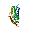 6zleC 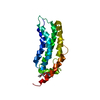 6zprC  6zvoC 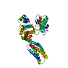 4btfS C: citing same article ( S: Starting model for refinement |
|---|---|
| Similar structure data |

-Links
 PDBj
PDBj

- Assembly
| Deposited unit | 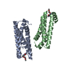
| ||||||||
|---|---|---|---|---|---|---|---|---|---|
| 1 | 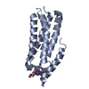
| ||||||||
| 2 | 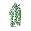
| ||||||||
| Unit cell |
|
-Components
| #1: Protein | Mass: 17642.326 Da / Num. of mol.: 2 Source method: isolated from a genetically manipulated source Source: (gene. exp.) Homo sapiens (human) / Gene: MLKL / Production host:  #2: Chemical | 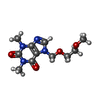  Mass: 268.269 Da / Num. of mol.: 2 / Source method: obtained synthetically / Formula: C11H16N4O4 / Feature type: SUBJECT OF INVESTIGATION Mass: 268.269 Da / Num. of mol.: 2 / Source method: obtained synthetically / Formula: C11H16N4O4 / Feature type: SUBJECT OF INVESTIGATION#3: Water | ChemComp-HOH / |  Mass: 18.015 Da / Num. of mol.: 154 / Source method: isolated from a natural source / Formula: H2O Mass: 18.015 Da / Num. of mol.: 154 / Source method: isolated from a natural source / Formula: H2OHas ligand of interest | Y | Has protein modification | Y | |
|---|
-Experimental details
-Experiment
| Experiment | Method: X-RAY DIFFRACTION / Number of used crystals: 1 |
|---|
- Sample preparation
| Crystal | Density Matthews: 2.06 Å3/Da / Density % sol: 40.35 % |
|---|---|
| Crystal grow | Temperature: 277 K / Method: vapor diffusion, hanging drop Details: 32 % Polyethylene glycol monomethyl ether 2,000, 0.15 M Potassium bromide, 0.1 M TRIS, pH 9 |
-Data collection
| Diffraction | Mean temperature: 100 K / Serial crystal experiment: N |
|---|---|
| Diffraction source | Source: SYNCHROTRON / Site: SLS  / Beamline: X10SA / Wavelength: 0.99994 Å / Beamline: X10SA / Wavelength: 0.99994 Å |
| Detector | Type: DECTRIS PILATUS 6M-F / Detector: PIXEL / Date: Sep 1, 2018 |
| Radiation | Protocol: SINGLE WAVELENGTH / Monochromatic (M) / Laue (L): M / Scattering type: x-ray |
| Radiation wavelength | Wavelength: 0.99994 Å / Relative weight: 1 |
| Reflection | Resolution: 1.64→19.705 Å / Num. obs: 28984 / % possible obs: 82.8 % / Redundancy: 6.7 % / Rmerge(I) obs: 0.029 / Rsym value: 0.029 / Net I/σ(I): 24.6 |
| Reflection shell | Resolution: 1.64→1.746 Å / Redundancy: 5.5 % / Rmerge(I) obs: 0.797 / Mean I/σ(I) obs: 1.4 / Num. unique obs: 1449 / Rsym value: 0.797 / % possible all: 24.3 |
- Processing
| Software |
| ||||||||||||||||||||||||||||||||||||||||||||||||||||||||||||
|---|---|---|---|---|---|---|---|---|---|---|---|---|---|---|---|---|---|---|---|---|---|---|---|---|---|---|---|---|---|---|---|---|---|---|---|---|---|---|---|---|---|---|---|---|---|---|---|---|---|---|---|---|---|---|---|---|---|---|---|---|---|
| Refinement | Method to determine structure: MOLECULAR REPLACEMENT Starting model: 4btf Resolution: 1.64→20.05 Å / Cor.coef. Fo:Fc: 0.956 / Cor.coef. Fo:Fc free: 0.951 / SU R Cruickshank DPI: 0.125 / Cross valid method: THROUGHOUT / SU R Blow DPI: 0.126 / SU Rfree Blow DPI: 0.118 / SU Rfree Cruickshank DPI: 0.119
| ||||||||||||||||||||||||||||||||||||||||||||||||||||||||||||
| Displacement parameters | Biso mean: 52.09 Å2
| ||||||||||||||||||||||||||||||||||||||||||||||||||||||||||||
| Refine analyze | Luzzati coordinate error obs: 0.24 Å | ||||||||||||||||||||||||||||||||||||||||||||||||||||||||||||
| Refinement step | Cycle: LAST / Resolution: 1.64→20.05 Å
| ||||||||||||||||||||||||||||||||||||||||||||||||||||||||||||
| Refine LS restraints |
| ||||||||||||||||||||||||||||||||||||||||||||||||||||||||||||
| LS refinement shell | Resolution: 1.64→1.7 Å
|
