 Movie
Movie Controller


+ Open data

- Basic information
| Entry | Database: PDB / ID: 6unj | ||||||
|---|---|---|---|---|---|---|---|
| Title | Human CYP3A4 bound to an inhibitor | ||||||
 Components Components | Cytochrome P450 3A4 | ||||||
 Keywords Keywords | OXIDOREDUCTASE/OXIDOREDUCTASE INHIBITOR / CYP3A4 / inhibitor / complex / OXIDOREDUCTASE / OXIDOREDUCTASE-OXIDOREDUCTASE INHIBITOR complex | ||||||
| Function / homology |  Function and homology information Function and homology informationquinine 3-monooxygenase / 1,8-cineole 2-exo-monooxygenase / albendazole monooxygenase (sulfoxide-forming) / quinine 3-monooxygenase activity / 1,8-cineole 2-exo-monooxygenase activity / 1-alpha,25-dihydroxyvitamin D3 23-hydroxylase activity / vitamin D 25-hydroxylase activity / vitamin D 24-hydroxylase activity / vitamin D catabolic process / retinoic acid 4-hydroxylase activity ...quinine 3-monooxygenase / 1,8-cineole 2-exo-monooxygenase / albendazole monooxygenase (sulfoxide-forming) / quinine 3-monooxygenase activity / 1,8-cineole 2-exo-monooxygenase activity / 1-alpha,25-dihydroxyvitamin D3 23-hydroxylase activity / vitamin D 25-hydroxylase activity / vitamin D 24-hydroxylase activity / vitamin D catabolic process / retinoic acid 4-hydroxylase activity / aflatoxin metabolic process / caffeine oxidase activity / estrogen 16-alpha-hydroxylase activity / lipid hydroxylation / anandamide 8,9 epoxidase activity / anandamide 11,12 epoxidase activity / anandamide 14,15 epoxidase activity / testosterone 6-beta-hydroxylase activity / alkaloid catabolic process / Aflatoxin activation and detoxification / Biosynthesis of maresin-like SPMs / monoterpenoid metabolic process / estrogen 2-hydroxylase activity / steroid catabolic process / oxidative demethylation / vitamin D metabolic process / Atorvastatin ADME / steroid hydroxylase activity / Xenobiotics / retinoic acid metabolic process / Phase I - Functionalization of compounds / estrogen metabolic process / long-chain fatty acid biosynthetic process / retinol metabolic process / unspecific monooxygenase / Prednisone ADME / Aspirin ADME / steroid metabolic process / androgen metabolic process / intracellular membrane-bounded organelle / cholesterol metabolic process / xenobiotic catabolic process / steroid binding / xenobiotic metabolic process / lipid metabolic process / monooxygenase activity / oxygen binding / oxidoreductase activity / iron ion binding / heme binding / endoplasmic reticulum membrane / enzyme binding / cytoplasm Similarity search - Function | ||||||
| Biological species |  Homo sapiens (human) Homo sapiens (human) | ||||||
| Method |  X-RAY DIFFRACTION / SYNCHROTRON / MOLECULAR REPLACEMENT / Resolution: 2.6 Å X-RAY DIFFRACTION / SYNCHROTRON / MOLECULAR REPLACEMENT / Resolution: 2.6 Å | ||||||
 Authors Authors | Sevrioukova, I. | ||||||
| Funding support |  United States, 1items United States, 1items
| ||||||
 Citation Citation | Journal: Bioorg.Med.Chem. / Year: 2020 Title: An increase in side-group hydrophobicity largely improves the potency of ritonavir-like inhibitors of CYP3A4. Authors: Samuels, E.R. / Sevrioukova, I.F. | ||||||
| History |
|
- Structure visualization
| Structure viewer | Molecule: MolmilJmol/JSmol |
|---|
- Downloads & links
-Download
| PDBx/mmCIF format | 6unj.cif.gz | 383.8 KB | Display | PDBx/mmCIF format |
|---|---|---|---|---|
| PDB format | pdb6unj.ent.gz | 317.2 KB | Display | PDB format |
| PDBx/mmJSON format | 6unj.json.gz | Tree view | PDBx/mmJSON format | |
| Others |  Other downloads Other downloads |
-Validation report
| Arichive directory | https://data.pdbj.org/pub/pdb/validation_reports/un/6unjftp://data.pdbj.org/pub/pdb/validation_reports/un/6unj | HTTPS FTP |
|---|
-Related structure data
| Related structure data | 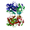 6uneC 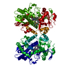 6ungC 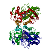 6unhC  6uniC 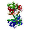 6unkC  6unlC  6unmC  5vccS C: citing same article ( S: Starting model for refinement |
|---|---|
| Similar structure data |


-Links
 PDBj
PDBj

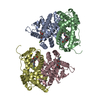
- Assembly
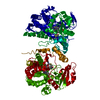
| Deposited unit | 
| ||||||||
|---|---|---|---|---|---|---|---|---|---|
| 1 | 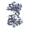
| ||||||||
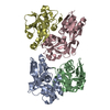
| 2 | 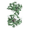
| ||||||||
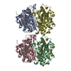
| 3 | 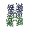
| ||||||||
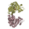
| Unit cell |
|
-Components
| #1: Protein | Mass: 55757.812 Da / Num. of mol.: 2 Source method: isolated from a genetically manipulated source Source: (gene. exp.) Homo sapiens (human) / Gene: CYP3A4, CYP3A3 / Production host:  References: UniProt: P08684, unspecific monooxygenase, 1,8-cineole 2-exo-monooxygenase, albendazole monooxygenase (sulfoxide-forming), quinine 3-monooxygenase #2: Chemical |   Mass: 616.487 Da / Num. of mol.: 2 / Source method: obtained synthetically / Formula: C34H32FeN4O4 Mass: 616.487 Da / Num. of mol.: 2 / Source method: obtained synthetically / Formula: C34H32FeN4O4#3: Chemical | 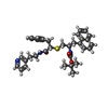  Mass: 569.757 Da / Num. of mol.: 2 / Source method: obtained synthetically / Formula: C34H39N3O3S / Feature type: SUBJECT OF INVESTIGATION Mass: 569.757 Da / Num. of mol.: 2 / Source method: obtained synthetically / Formula: C34H39N3O3S / Feature type: SUBJECT OF INVESTIGATIONHas ligand of interest | Y | |
|---|
-Experimental details
-Experiment
| Experiment | Method: X-RAY DIFFRACTION / Number of used crystals: 1 |
|---|
- Sample preparation
| Crystal | Density Matthews: 2.53 Å3/Da / Density % sol: 51.37 % |
|---|---|
| Crystal grow | Temperature: 298 K / Method: microbatch / Details: PEG3350, sodium malonate / PH range: 6.0-7.0 |
-Data collection
| Diffraction | Mean temperature: 77 K / Serial crystal experiment: N | |||||||||||||||||||||||||||
|---|---|---|---|---|---|---|---|---|---|---|---|---|---|---|---|---|---|---|---|---|---|---|---|---|---|---|---|---|
| Diffraction source | Source: SYNCHROTRON / Site: SSRL / Beamline: BL9-2 / Wavelength: 0.98 Å | |||||||||||||||||||||||||||
| Detector | Type: DECTRIS PILATUS 6M / Detector: PIXEL / Date: May 18, 2018 / Details: mirrors | |||||||||||||||||||||||||||
| Radiation | Protocol: SINGLE WAVELENGTH / Monochromatic (M) / Laue (L): M / Scattering type: x-ray | |||||||||||||||||||||||||||
| Radiation wavelength | Wavelength: 0.98 Å / Relative weight: 1 | |||||||||||||||||||||||||||
| Reflection | Resolution: 2.6→77.57 Å / Num. obs: 32836 / % possible obs: 93.2 % / Redundancy: 6 % / Biso Wilson estimate: 91.46 Å2 / CC1/2: 0.999 / Rmerge(I) obs: 0.083 / Rpim(I) all: 0.036 / Rrim(I) all: 0.091 / Net I/σ(I): 7.6 / Num. measured all: 196720 | |||||||||||||||||||||||||||
| Reflection shell | Diffraction-ID: 1 / Redundancy: 6 %
|
- Processing
| Software |
| ||||||||||||||||||||||||||||||||||||||||||||||||||||||||||||||||||||||||||||||
|---|---|---|---|---|---|---|---|---|---|---|---|---|---|---|---|---|---|---|---|---|---|---|---|---|---|---|---|---|---|---|---|---|---|---|---|---|---|---|---|---|---|---|---|---|---|---|---|---|---|---|---|---|---|---|---|---|---|---|---|---|---|---|---|---|---|---|---|---|---|---|---|---|---|---|---|---|---|---|---|
| Refinement | Method to determine structure: MOLECULAR REPLACEMENT Starting model: 5VCC Resolution: 2.6→47.294 Å / SU ML: 0.57 / Cross valid method: THROUGHOUT / σ(F): 1.33 / Phase error: 45.1
| ||||||||||||||||||||||||||||||||||||||||||||||||||||||||||||||||||||||||||||||
| Solvent computation | Shrinkage radii: 0.9 Å / VDW probe radii: 1.11 Å | ||||||||||||||||||||||||||||||||||||||||||||||||||||||||||||||||||||||||||||||
| Displacement parameters | Biso max: 339.16 Å2 / Biso mean: 147.5553 Å2 / Biso min: 48.32 Å2 | ||||||||||||||||||||||||||||||||||||||||||||||||||||||||||||||||||||||||||||||
| Refinement step | Cycle: final / Resolution: 2.6→47.294 Å
| ||||||||||||||||||||||||||||||||||||||||||||||||||||||||||||||||||||||||||||||
| Refine LS restraints |
| ||||||||||||||||||||||||||||||||||||||||||||||||||||||||||||||||||||||||||||||
| LS refinement shell | Refine-ID: X-RAY DIFFRACTION / Rfactor Rfree error: 0
| ||||||||||||||||||||||||||||||||||||||||||||||||||||||||||||||||||||||||||||||
| Refinement TLS params. | Method: refined / Origin x: -27.7675 Å / Origin y: -25.5309 Å / Origin z: 108.6095 Å
| ||||||||||||||||||||||||||||||||||||||||||||||||||||||||||||||||||||||||||||||
| Refinement TLS group |
|
