 Movie
Movie Controller


+ Open data

- Basic information
| Entry | Database: PDB / ID: 3t22 | ||||||
|---|---|---|---|---|---|---|---|
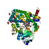
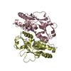
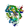
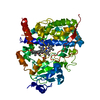
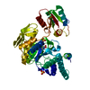
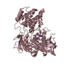
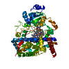
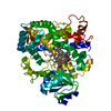
| Title | Crystal structure of OxyR mutant from Porphyromonas gingivalis | ||||||
 Components Components | Redox-sensitive transcriptional activator OxyR | ||||||
 Keywords Keywords | TRANSCRIPTION REGULATOR / beta-alpha-barrels / DNA-binding / Transcription regulation | ||||||
| Function / homology |  Function and homology information Function and homology informationDNA-binding transcription factor activity / DNA-templated transcription / DNA binding / cytosol Similarity search - Function | ||||||
| Biological species |  Porphyromonas gingivalis (bacteria) Porphyromonas gingivalis (bacteria) | ||||||
| Method |  X-RAY DIFFRACTION / MOLECULAR REPLACEMENT / Resolution: 2.2 Å X-RAY DIFFRACTION / MOLECULAR REPLACEMENT / Resolution: 2.2 Å | ||||||
 Authors Authors | Svintradze, D.V. / Wright, H.T. / Lewis, J.P. | ||||||
 Citation Citation | Journal: Acta Crystallogr.,Sect.D / Year: 2013 Title: Structures of the Porphyromonas gingivalis OxyR regulatory domain explain differences in expression of the OxyR regulon in Escherichia coli and P. gingivalis. Authors: Svintradze, D.V. / Peterson, D.L. / Collazo-Santiago, E.A. / Lewis, J.P. / Wright, H.T. #1: Journal: To be PublishedTitle: The iron-oxygen molecular switch in the anaerobic bacterium Porphyromonas gingivalis Authors: Lewis, J.P. / Yanamandra, S. / Ghosh, A.K. / Svintradze, D.V. / Sengupta, D. / Jones, K. / Iyer, D. / Peterson, D. / Wright, H.T. / Anaya-Bergman, C. | ||||||
| History |
|
- Structure visualization
| Structure viewer | Molecule: MolmilJmol/JSmol |
|---|
- Downloads & links
-Download
| PDBx/mmCIF format | 3t22.cif.gz | 181.8 KB | Display | PDBx/mmCIF format |
|---|---|---|---|---|
| PDB format | pdb3t22.ent.gz | 145.7 KB | Display | PDB format |
| PDBx/mmJSON format | 3t22.json.gz | Tree view | PDBx/mmJSON format | |
| Others |  Other downloads Other downloads |
-Validation report
| Arichive directory | https://data.pdbj.org/pub/pdb/validation_reports/t2/3t22ftp://data.pdbj.org/pub/pdb/validation_reports/t2/3t22 | HTTPS FTP |
|---|
-Related structure data
| Related structure data |  3ho7SC  3ukiC S: Starting model for refinement C: citing same article ( |
|---|---|
| Similar structure data |


-Links
 PDBj
PDBj- Assembly
| Deposited unit | 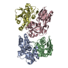
| ||||||||
|---|---|---|---|---|---|---|---|---|---|
| 1 | 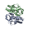
| ||||||||
| 2 | 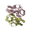
| ||||||||
| Unit cell |
|
-Components
| #1: Protein | Mass: 25605.510 Da / Num. of mol.: 4 / Fragment: Regulatory domain (UNP residues 90-308) / Mutation: C199S Source method: isolated from a genetically manipulated source Source: (gene. exp.) Porphyromonas gingivalis (bacteria) / Gene: oxyR / Production host: #2: Water | ChemComp-HOH / |  Mass: 18.015 Da / Num. of mol.: 378 / Source method: isolated from a natural source / Formula: H2O Mass: 18.015 Da / Num. of mol.: 378 / Source method: isolated from a natural source / Formula: H2O |
|---|
-Experimental details
-Experiment
| Experiment | Method: X-RAY DIFFRACTION / Number of used crystals: 1 |
|---|
- Sample preparation
| Crystal | Density Matthews: 2.45 Å3/Da / Density % sol: 49.83 % |
|---|---|
| Crystal grow | Temperature: 293 K / Method: vapor diffusion, sitting drop / pH: 9.5 Details: 30% PEG 3000, 0.1M CHES, pH 9.5, VAPOR DIFFUSION, SITTING DROP, temperature 293K |
-Data collection
| Diffraction | Mean temperature: 100 K |
|---|---|
| Diffraction source | Source: ROTATING ANODE / Type: RIGAKU MICROMAX-007 / Wavelength: 1.5418 Å |
| Detector | Type: RIGAKU RAXIS IV++ / Detector: IMAGE PLATE / Date: Jun 23, 2011 |
| Radiation | Monochromator: MSC Varimax confocal optics / Protocol: SINGLE WAVELENGTH / Monochromatic (M) / Laue (L): M / Scattering type: x-ray |
| Radiation wavelength | Wavelength: 1.5418 Å / Relative weight: 1 |
| Reflection | Resolution: 1.9→23.24 Å / Num. obs: 69702 / % possible obs: 90.9 % / Observed criterion σ(F): 0 / Observed criterion σ(I): 3 / Redundancy: 3.53 % / Rmerge(I) obs: 0.1 / Net I/σ(I): 5.8 |
| Reflection shell | Resolution: 1.9→1.97 Å / Redundancy: 3.32 % / Rmerge(I) obs: 0.794 / Mean I/σ(I) obs: 0.9 / % possible all: 88.8 |
- Processing
| Software |
| |||||||||||||||||||||||||||||||||||||||||||||||||||||||||||||||||||||||||||||
|---|---|---|---|---|---|---|---|---|---|---|---|---|---|---|---|---|---|---|---|---|---|---|---|---|---|---|---|---|---|---|---|---|---|---|---|---|---|---|---|---|---|---|---|---|---|---|---|---|---|---|---|---|---|---|---|---|---|---|---|---|---|---|---|---|---|---|---|---|---|---|---|---|---|---|---|---|---|---|
| Refinement | Method to determine structure: MOLECULAR REPLACEMENT Starting model: 3HO7 Resolution: 2.2→18.358 Å / SU ML: 0.39 / σ(F): 0 / Phase error: 31.7 / Stereochemistry target values: ML Details: The structure factor file contains 69444 reflections with the resolution range of 1.90-23.24 A. However the resolution of 2.2-18.358 A with 44680 reflections was used for the refinement. The ...Details: The structure factor file contains 69444 reflections with the resolution range of 1.90-23.24 A. However the resolution of 2.2-18.358 A with 44680 reflections was used for the refinement. The decision of cutting the high resolution reflections was based on the low intensity signals for 1.90 A resolution which were keeping R factor high during the refinement.
| |||||||||||||||||||||||||||||||||||||||||||||||||||||||||||||||||||||||||||||
| Solvent computation | Shrinkage radii: 0.95 Å / VDW probe radii: 1.2 Å / Solvent model: FLAT BULK SOLVENT MODEL / Bsol: 40.001 Å2 / ksol: 0.344 e/Å3 | |||||||||||||||||||||||||||||||||||||||||||||||||||||||||||||||||||||||||||||
| Displacement parameters |
| |||||||||||||||||||||||||||||||||||||||||||||||||||||||||||||||||||||||||||||
| Refinement step | Cycle: LAST / Resolution: 2.2→18.358 Å
| |||||||||||||||||||||||||||||||||||||||||||||||||||||||||||||||||||||||||||||
| Refine LS restraints |
| |||||||||||||||||||||||||||||||||||||||||||||||||||||||||||||||||||||||||||||
| LS refinement shell |
|
