 Movie
Movie Controller


+ Open data

- Basic information
| Entry | Database: PDB / ID: 4d78 | ||||||
|---|---|---|---|---|---|---|---|
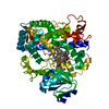
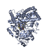
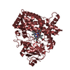
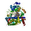
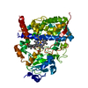
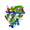
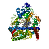
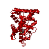
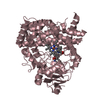
| Title | Cytochrome P450 3A4 bound to an inhibitor | ||||||
 Components Components | CYTOCHROME P450 3A4 | ||||||
 Keywords Keywords | OXIDOREDUCTASE / MONOOXYGENASE / HUMAN CYP3A4 / INHIBITORY COMPLEX | ||||||
| Function / homology |  Function and homology information Function and homology informationquinine 3-monooxygenase / 1,8-cineole 2-exo-monooxygenase / albendazole monooxygenase (sulfoxide-forming) / quinine 3-monooxygenase activity / 1,8-cineole 2-exo-monooxygenase activity / 1-alpha,25-dihydroxyvitamin D3 23-hydroxylase activity / vitamin D 25-hydroxylase activity / vitamin D 24-hydroxylase activity / vitamin D catabolic process / retinoic acid 4-hydroxylase activity ...quinine 3-monooxygenase / 1,8-cineole 2-exo-monooxygenase / albendazole monooxygenase (sulfoxide-forming) / quinine 3-monooxygenase activity / 1,8-cineole 2-exo-monooxygenase activity / 1-alpha,25-dihydroxyvitamin D3 23-hydroxylase activity / vitamin D 25-hydroxylase activity / vitamin D 24-hydroxylase activity / vitamin D catabolic process / retinoic acid 4-hydroxylase activity / caffeine oxidase activity / estrogen 16-alpha-hydroxylase activity / lipid hydroxylation / aflatoxin metabolic process / anandamide 8,9 epoxidase activity / anandamide 11,12 epoxidase activity / anandamide 14,15 epoxidase activity / testosterone 6-beta-hydroxylase activity / alkaloid catabolic process / Aflatoxin activation and detoxification / Biosynthesis of maresin-like SPMs / monoterpenoid metabolic process / estrogen 2-hydroxylase activity / steroid catabolic process / oxidative demethylation / vitamin D metabolic process / Atorvastatin ADME / steroid hydroxylase activity / Xenobiotics / retinoic acid metabolic process / Phase I - Functionalization of compounds / estrogen metabolic process / retinol metabolic process / unspecific monooxygenase / long-chain fatty acid biosynthetic process / Prednisone ADME / Aspirin ADME / steroid metabolic process / androgen metabolic process / intracellular membrane-bounded organelle / xenobiotic catabolic process / cholesterol metabolic process / steroid binding / xenobiotic metabolic process / lipid metabolic process / monooxygenase activity / oxygen binding / oxidoreductase activity / iron ion binding / heme binding / endoplasmic reticulum membrane / enzyme binding / cytoplasm Similarity search - Function | ||||||
| Biological species |  HOMO SAPIENS (human) HOMO SAPIENS (human) | ||||||
| Method |  X-RAY DIFFRACTION / SYNCHROTRON / MOLECULAR REPLACEMENT / Resolution: 2.8 Å X-RAY DIFFRACTION / SYNCHROTRON / MOLECULAR REPLACEMENT / Resolution: 2.8 Å | ||||||
 Authors Authors | Sevrioukova, I. / Poulos, T. | ||||||
 Citation Citation | Journal: J.Med.Chem. / Year: 2016 Title: Structure-Based Inhibitor Design for Evaluation of a Cyp3A4 Pharmacophore Model. Authors: Kaur, P. / Chamberlin, R. / Poulos, T.L. / Sevrioukova, I.F. | ||||||
| History |
|
- Structure visualization
| Structure viewer | Molecule: MolmilJmol/JSmol |
|---|
- Downloads & links
-Download
| PDBx/mmCIF format | 4d78.cif.gz | 205.4 KB | Display | PDBx/mmCIF format |
|---|---|---|---|---|
| PDB format | pdb4d78.ent.gz | 164.2 KB | Display | PDB format |
| PDBx/mmJSON format | 4d78.json.gz | Tree view | PDBx/mmJSON format | |
| Others |  Other downloads Other downloads |
-Validation report
| Arichive directory | https://data.pdbj.org/pub/pdb/validation_reports/d7/4d78ftp://data.pdbj.org/pub/pdb/validation_reports/d7/4d78 | HTTPS FTP |
|---|
-Related structure data
| Related structure data | 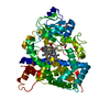 4d6zC  4d75C  4d7dC  1tqnS C: citing same article ( S: Starting model for refinement |
|---|---|
| Similar structure data |

-Links
 PDBj
PDBj

- Assembly
| Deposited unit | 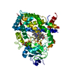
| ||||||||
|---|---|---|---|---|---|---|---|---|---|
| 1 |
| ||||||||
| Unit cell |
|
-Components
| #1: Protein | Mass: 55757.812 Da / Num. of mol.: 1 / Fragment: CATALYTIC DOMAIN, UNP RESIDUES 23-503 Source method: isolated from a genetically manipulated source Source: (gene. exp.) HOMO SAPIENS (human) / Plasmid: PCWORI / Production host:  References: UniProt: P08684, EC: 1.14.13.157, EC: 1.14.13.32, EC: 1.14.13.67, EC: 1.14.13.97 | ||||
|---|---|---|---|---|---|
| #2: Chemical | ChemComp-HEM /   Mass: 616.487 Da / Num. of mol.: 1 / Source method: obtained synthetically / Formula: C34H32FeN4O4 Mass: 616.487 Da / Num. of mol.: 1 / Source method: obtained synthetically / Formula: C34H32FeN4O4 | ||||
| #3: Chemical | ChemComp-J9K /   Mass: 429.576 Da / Num. of mol.: 1 / Source method: obtained synthetically / Formula: C23H31N3O3S Mass: 429.576 Da / Num. of mol.: 1 / Source method: obtained synthetically / Formula: C23H31N3O3S | ||||
| #4: Chemical |   Mass: 92.094 Da / Num. of mol.: 2 / Source method: obtained synthetically / Formula: C3H8O3 Mass: 92.094 Da / Num. of mol.: 2 / Source method: obtained synthetically / Formula: C3H8O3#5: Water | ChemComp-HOH / |  Mass: 18.015 Da / Num. of mol.: 6 / Source method: isolated from a natural source / Formula: H2O Mass: 18.015 Da / Num. of mol.: 6 / Source method: isolated from a natural source / Formula: H2ONonpolymer details | NEEDS TO BE NAMED (LIG): FIGURE WITH THE HETEROGROU | |
-Experimental details
-Experiment
| Experiment | Method: X-RAY DIFFRACTION / Number of used crystals: 1 |
|---|
- Sample preparation
| Crystal | Density Matthews: 2.38 Å3/Da / Density % sol: 48.3 % / Description: NONE |
|---|---|
| Crystal grow | Method: microbatch / pH: 5 Details: MICROBATCH UNDER OIL WITH 10% PEG 3350 AND 80 MM SODIUM MALONATE PH 5.0 |
-Data collection
| Diffraction | Mean temperature: 113 K |
|---|---|
| Diffraction source | Source: SYNCHROTRON / Site: ALS  / Beamline: 8.2.1 / Wavelength: 1 / Beamline: 8.2.1 / Wavelength: 1 |
| Detector | Type: ADSC CCD / Detector: CCD / Date: Apr 24, 2013 / Details: MIRRORS |
| Radiation | Protocol: SINGLE WAVELENGTH / Monochromatic (M) / Laue (L): M / Scattering type: x-ray |
| Radiation wavelength | Wavelength: 1 Å / Relative weight: 1 |
| Reflection | Resolution: 2.8→65.9 Å / Num. obs: 12092 / % possible obs: 100 % / Observed criterion σ(I): 2 / Redundancy: 5.6 % / Rmerge(I) obs: 0.08 / Net I/σ(I): 10.6 |
| Reflection shell | Resolution: 2.8→2.95 Å / Redundancy: 5.9 % / Rmerge(I) obs: 0.4 / Mean I/σ(I) obs: 1.9 / % possible all: 100 |
- Processing
| Software |
| ||||||||||||||||||||||||||||||||||||||||||||||||||||||||||||||||||||||||||||||||||||||||||||||||||||||||||||||||||||||||||||||||||||||||||||||||||||||||||||||||||||||||||||||||||||||
|---|---|---|---|---|---|---|---|---|---|---|---|---|---|---|---|---|---|---|---|---|---|---|---|---|---|---|---|---|---|---|---|---|---|---|---|---|---|---|---|---|---|---|---|---|---|---|---|---|---|---|---|---|---|---|---|---|---|---|---|---|---|---|---|---|---|---|---|---|---|---|---|---|---|---|---|---|---|---|---|---|---|---|---|---|---|---|---|---|---|---|---|---|---|---|---|---|---|---|---|---|---|---|---|---|---|---|---|---|---|---|---|---|---|---|---|---|---|---|---|---|---|---|---|---|---|---|---|---|---|---|---|---|---|---|---|---|---|---|---|---|---|---|---|---|---|---|---|---|---|---|---|---|---|---|---|---|---|---|---|---|---|---|---|---|---|---|---|---|---|---|---|---|---|---|---|---|---|---|---|---|---|---|---|
| Refinement | Method to determine structure: MOLECULAR REPLACEMENT Starting model: PDB ENTRY 1TQN Resolution: 2.8→64.85 Å / Cor.coef. Fo:Fc: 0.951 / Cor.coef. Fo:Fc free: 0.9 / SU B: 41.476 / SU ML: 0.37 / Cross valid method: THROUGHOUT / ESU R Free: 0.446 / Stereochemistry target values: MAXIMUM LIKELIHOOD Details: HYDROGENS HAVE BEEN ADDED IN THE RIDING POSITIONS. RESIDUES 265-266 AND 280-288 ARE DISORDERED
| ||||||||||||||||||||||||||||||||||||||||||||||||||||||||||||||||||||||||||||||||||||||||||||||||||||||||||||||||||||||||||||||||||||||||||||||||||||||||||||||||||||||||||||||||||||||
| Solvent computation | Ion probe radii: 0.8 Å / Shrinkage radii: 0.8 Å / VDW probe radii: 1.2 Å / Solvent model: MASK | ||||||||||||||||||||||||||||||||||||||||||||||||||||||||||||||||||||||||||||||||||||||||||||||||||||||||||||||||||||||||||||||||||||||||||||||||||||||||||||||||||||||||||||||||||||||
| Displacement parameters | Biso mean: 102.012 Å2
| ||||||||||||||||||||||||||||||||||||||||||||||||||||||||||||||||||||||||||||||||||||||||||||||||||||||||||||||||||||||||||||||||||||||||||||||||||||||||||||||||||||||||||||||||||||||
| Refinement step | Cycle: LAST / Resolution: 2.8→64.85 Å
| ||||||||||||||||||||||||||||||||||||||||||||||||||||||||||||||||||||||||||||||||||||||||||||||||||||||||||||||||||||||||||||||||||||||||||||||||||||||||||||||||||||||||||||||||||||||
| Refine LS restraints |
|