 Movie
Movie Controller


+ Open data

- Basic information
| Entry | Database: PDB / ID: 3h45 | ||||||
|---|---|---|---|---|---|---|---|
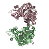
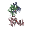
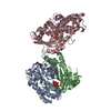
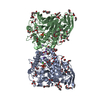
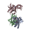
| Title | Glycerol Kinase H232E with Ethylene Glycol | ||||||
 Components Components | Glycerol kinase | ||||||
 Keywords Keywords | TRANSFERASE / ethylene glycol / kinase / ATP-binding / Glycerol metabolism / Nucleotide-binding / Phosphoprotein | ||||||
| Function / homology |  Function and homology information Function and homology informationglycerol-3-phosphate metabolic process / glycerol kinase / glycerol kinase activity / glycerol metabolic process / glycerol catabolic process / ATP binding / cytosol Similarity search - Function | ||||||
| Biological species |  Enterococcus casseliflavus (bacteria) Enterococcus casseliflavus (bacteria) | ||||||
| Method |  X-RAY DIFFRACTION / MOLECULAR REPLACEMENT / Resolution: 2.65 Å X-RAY DIFFRACTION / MOLECULAR REPLACEMENT / Resolution: 2.65 Å | ||||||
 Authors Authors | Yeh, J.I. / Kettering, R.D. | ||||||
 Citation Citation | Journal: Biochemistry / Year: 2009 Title: Structural characterizations of glycerol kinase: unraveling phosphorylation-induced long-range activation Authors: Yeh, J.I. / Kettering, R. / Saxl, R. / Bourand, A. / Darbon, E. / Joly, N. / Briozzo, P. / Deutscher, J. | ||||||
| History |
|
- Structure visualization
| Structure viewer | Molecule: MolmilJmol/JSmol |
|---|
- Downloads & links
-Download
| PDBx/mmCIF format | 3h45.cif.gz | 389.3 KB | Display | PDBx/mmCIF format |
|---|---|---|---|---|
| PDB format | pdb3h45.ent.gz | 317.9 KB | Display | PDB format |
| PDBx/mmJSON format | 3h45.json.gz | Tree view | PDBx/mmJSON format | |
| Others |  Other downloads Other downloads |
-Validation report
| Arichive directory | https://data.pdbj.org/pub/pdb/validation_reports/h4/3h45ftp://data.pdbj.org/pub/pdb/validation_reports/h4/3h45 | HTTPS FTP |
|---|
-Related structure data
| Related structure data |  3d7eC  3flcC  3h3nC  3h3oC  3h46C  1xupS C: citing same article ( S: Starting model for refinement |
|---|---|
| Similar structure data |


-Links
 PDBj
PDBj- Assembly
| Deposited unit | 
| ||||||||
|---|---|---|---|---|---|---|---|---|---|
| 1 | 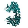
| ||||||||
| 2 | 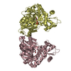
| ||||||||
| 3 | 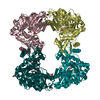
| ||||||||
| Unit cell |
|
-Components
| #1: Protein | Mass: 55803.453 Da / Num. of mol.: 4 / Mutation: H232E Source method: isolated from a genetically manipulated source Source: (gene. exp.) Enterococcus casseliflavus (bacteria) / Gene: glpK / Plasmid: pOXO4 / Production host: #2: Chemical | ChemComp-EDO /   Mass: 62.068 Da / Num. of mol.: 5 / Source method: obtained synthetically / Formula: C2H6O2 Mass: 62.068 Da / Num. of mol.: 5 / Source method: obtained synthetically / Formula: C2H6O2#3: Chemical | ChemComp-PO4 /   Mass: 94.971 Da / Num. of mol.: 6 / Source method: obtained synthetically / Formula: PO4 Mass: 94.971 Da / Num. of mol.: 6 / Source method: obtained synthetically / Formula: PO4#4: Water | ChemComp-HOH / |  Mass: 18.015 Da / Num. of mol.: 300 / Source method: isolated from a natural source / Formula: H2O Mass: 18.015 Da / Num. of mol.: 300 / Source method: isolated from a natural source / Formula: H2O |
|---|
-Experimental details
-Experiment
| Experiment | Method: X-RAY DIFFRACTION / Number of used crystals: 1 |
|---|
- Sample preparation
| Crystal | Density Matthews: 2.42 Å3/Da / Density % sol: 49.08 % |
|---|---|
| Crystal grow | Temperature: 298 K / Method: vapor diffusion Details: KH2PO4, PEG 8000, VAPOR DIFFUSION, temperature 298K |
-Data collection
| Diffraction | Mean temperature: 93 K |
|---|---|
| Diffraction source | Source: ROTATING ANODE / Type: RIGAKU FR-E SUPERBRIGHT / Wavelength: 1.514 Å |
| Detector | Type: RIGAKU AFC11-KAPPA / Detector: DIFFRACTOMETER / Date: Jun 8, 2007 |
| Radiation | Protocol: SINGLE WAVELENGTH / Monochromatic (M) / Laue (L): M / Scattering type: x-ray |
| Radiation wavelength | Wavelength: 1.514 Å / Relative weight: 1 |
| Reflection | Resolution: 2.5→50 Å / Num. obs: 73411 / % possible obs: 99.8 % / Observed criterion σ(F): 0 / Observed criterion σ(I): 0 / Redundancy: 2.9 % / Rsym value: 0.09 / Net I/σ(I): 6.5 |
| Reflection shell | Resolution: 2.5→2.59 Å / Redundancy: 2.9 % / Mean I/σ(I) obs: 1.7 / Rsym value: 0.368 / % possible all: 99.8 |
- Processing
| Software |
| ||||||||||||||||||||||||||||
|---|---|---|---|---|---|---|---|---|---|---|---|---|---|---|---|---|---|---|---|---|---|---|---|---|---|---|---|---|---|
| Refinement | Method to determine structure: MOLECULAR REPLACEMENT Starting model: PDB ENTRY 1XUP Resolution: 2.65→12 Å / Occupancy max: 1 / Occupancy min: 1 / σ(F): 3 Stereochemistry target values: MAXIMUM LIKELIHOOD WITH PHASES
| ||||||||||||||||||||||||||||
| Displacement parameters | Biso max: 106.87 Å2 / Biso mean: 40.25 Å2 / Biso min: 8.47 Å2 | ||||||||||||||||||||||||||||
| Refinement step | Cycle: LAST / Resolution: 2.65→12 Å
| ||||||||||||||||||||||||||||
| Refine LS restraints |
|
