 Movie
Movie Controller

[English] 日本語
 Yorodumi
- PDB-3g82: Complex of GS-alpha with the catalytic domains of mammalian adeny... -
+ Open data

- Basic information
| Entry | Database: PDB / ID: 3g82 | ||||||
|---|---|---|---|---|---|---|---|
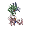
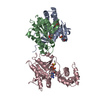
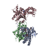
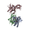
| Title | Complex of GS-alpha with the catalytic domains of mammalian adenylyl cyclase: complex with MANT-ITP and Mn | ||||||
 Components Components |
| ||||||
 Keywords Keywords | Lyase/Lyase Inhibitor / ADENYLYL CYCLASE / MANT-ITP / Alternative splicing / cAMP biosynthesis / Glycoprotein / Lyase / Magnesium / Membrane / Metal-binding / Phosphoprotein / Transmembrane / Cell membrane / GTP-binding / Lipoprotein / Nucleotide-binding / Palmitate / Transducer / Lyase-Lyase Inhibitor COMPLEX | ||||||
| Function / homology |  Function and homology information Function and homology informationAdenylate cyclase activating pathway / PKA activation / Hedgehog 'off' state / Adenylate cyclase inhibitory pathway / adenylate cyclase / regulation of insulin secretion involved in cellular response to glucose stimulus / sensory perception of chemical stimulus / mu-type opioid receptor binding / corticotropin-releasing hormone receptor 1 binding / adenylate cyclase activity ...Adenylate cyclase activating pathway / PKA activation / Hedgehog 'off' state / Adenylate cyclase inhibitory pathway / adenylate cyclase / regulation of insulin secretion involved in cellular response to glucose stimulus / sensory perception of chemical stimulus / mu-type opioid receptor binding / corticotropin-releasing hormone receptor 1 binding / adenylate cyclase activity / G alpha (z) signalling events / cAMP biosynthetic process / beta-2 adrenergic receptor binding / adenylate cyclase binding / D1 dopamine receptor binding / adenylate cyclase-activating adrenergic receptor signaling pathway / insulin-like growth factor receptor binding / ionotropic glutamate receptor binding / cellular response to forskolin / adenylate cyclase activator activity / G-protein beta/gamma-subunit complex binding / adenylate cyclase-modulating G protein-coupled receptor signaling pathway / manganese ion binding / adenylate cyclase-activating dopamine receptor signaling pathway / heterotrimeric G-protein complex / positive regulation of cytosolic calcium ion concentration / adenylate cyclase-activating G protein-coupled receptor signaling pathway / Hydrolases; Acting on acid anhydrides; Acting on GTP to facilitate cellular and subcellular movement / cilium / intracellular signal transduction / membrane raft / GTPase activity / dendrite / GTP binding / magnesium ion binding / protein-containing complex / ATP binding / membrane / metal ion binding / plasma membrane / cytoplasm Similarity search - Function | ||||||
| Biological species |  | ||||||
| Method |  X-RAY DIFFRACTION / SYNCHROTRON / MOLECULAR REPLACEMENT / Resolution: 3.11 Å X-RAY DIFFRACTION / SYNCHROTRON / MOLECULAR REPLACEMENT / Resolution: 3.11 Å | ||||||
 Authors Authors | Huebner, M. / Mou, T.-C. / Sprang, S.R. / Seifert, R. | ||||||
 Citation Citation | Journal: To be Published Title: 2',3'-(O)-(N-Methyl)anthraniloyl-inosine 5'-triphosphate is the Most Potent Adenylyl Cyclase 1 and 5 Inhibitor Known so far and Effectively Promotes Catalytic Subunit Assembly in the Absence of Forskolin Authors: Huebner, M. / Geduhn, J. / Pinto, C. / Mou, T.-C. / Konig, B. / Sprang, S.R. / Seifert, R. | ||||||
| History |
|
- Structure visualization
| Structure viewer | Molecule: MolmilJmol/JSmol |
|---|
- Downloads & links
-Download
| PDBx/mmCIF format | 3g82.cif.gz | 164.3 KB | Display | PDBx/mmCIF format |
|---|---|---|---|---|
| PDB format | pdb3g82.ent.gz | 123.9 KB | Display | PDB format |
| PDBx/mmJSON format | 3g82.json.gz | Tree view | PDBx/mmJSON format | |
| Others |  Other downloads Other downloads |
-Validation report
| Arichive directory | https://data.pdbj.org/pub/pdb/validation_reports/g8/3g82ftp://data.pdbj.org/pub/pdb/validation_reports/g8/3g82 | HTTPS FTP |
|---|
-Related structure data
| Related structure data |  1tl7S S: Starting model for refinement |
|---|---|
| Similar structure data |


-Links
 PDBj
PDBj


- Assembly
| Deposited unit | 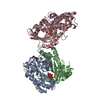
| ||||||||
|---|---|---|---|---|---|---|---|---|---|
| 1 |
| ||||||||
| Unit cell |
|
-Components
-Adenylate cyclase type ... , 2 types, 2 molecules AB
| #1: Protein | Mass: 25526.514 Da / Num. of mol.: 1 / Fragment: C1A DOMAIN / Mutation: V476M Source method: isolated from a genetically manipulated source Source: (gene. exp.)  |
|---|---|
| #2: Protein | Mass: 23717.033 Da / Num. of mol.: 1 / Fragment: C2A DOMAIN Source method: isolated from a genetically manipulated source Source: (gene. exp.) |
-Protein , 1 types, 1 molecules C
| #3: Protein | Mass: 45769.527 Da / Num. of mol.: 1 / Fragment: TRYPSINIZED FRAGMENT OF G(S)ALPHA SUBUNIT Source method: isolated from a genetically manipulated source Source: (gene. exp.) |
|---|
-Non-polymers , 7 types, 11 molecules 


| #4: Chemical |  Mass: 54.938 Da / Num. of mol.: 2 / Source method: obtained synthetically / Formula: Mn Mass: 54.938 Da / Num. of mol.: 2 / Source method: obtained synthetically / Formula: Mn#5: Chemical | ChemComp-FOK / |  Mass: 410.501 Da / Num. of mol.: 1 / Source method: obtained synthetically / Formula: C22H34O7 Mass: 410.501 Da / Num. of mol.: 1 / Source method: obtained synthetically / Formula: C22H34O7#6: Chemical | ChemComp-MI3 / |  Mass: 641.313 Da / Num. of mol.: 1 / Source method: obtained synthetically / Formula: C18H22N5O15P3 Mass: 641.313 Da / Num. of mol.: 1 / Source method: obtained synthetically / Formula: C18H22N5O15P3#7: Chemical | ChemComp-MG / |  Mass: 24.305 Da / Num. of mol.: 1 / Source method: obtained synthetically / Formula: Mg Mass: 24.305 Da / Num. of mol.: 1 / Source method: obtained synthetically / Formula: Mg#8: Chemical | ChemComp-CL / |  Mass: 35.453 Da / Num. of mol.: 1 / Source method: obtained synthetically / Formula: Cl Mass: 35.453 Da / Num. of mol.: 1 / Source method: obtained synthetically / Formula: Cl#9: Chemical | ChemComp-GSP / |  Mass: 539.246 Da / Num. of mol.: 1 / Source method: obtained synthetically / Formula: C10H16N5O13P3S Mass: 539.246 Da / Num. of mol.: 1 / Source method: obtained synthetically / Formula: C10H16N5O13P3S#10: Water | ChemComp-HOH / | Mass: 18.015 Da / Num. of mol.: 4 / Source method: isolated from a natural source / Formula: H2O |
|---|
-Experimental details
-Experiment
| Experiment | Method: X-RAY DIFFRACTION / Number of used crystals: 1 |
|---|
- Sample preparation
| Crystal | Density Matthews: 2.92 Å3/Da / Density % sol: 57.84 % |
|---|---|
| Crystal grow | Temperature: 289 K / Method: vapor diffusion, sitting drop / pH: 5.4 Details: 7.2-7.8% PEG8000, 0.5M NACL, 0.1M 2-morpholinoethanesulfonic acid, pH 5.4, VAPOR DIFFUSION, SITTING DROP, temperature 289K |
-Data collection
| Diffraction | Mean temperature: 100 K |
|---|---|
| Diffraction source | Source: SYNCHROTRON / Site: SSRL  / Beamline: BL9-1 / Wavelength: 0.97946 Å / Beamline: BL9-1 / Wavelength: 0.97946 Å |
| Detector | Type: ADSC QUANTUM 315 / Detector: CCD / Date: Jun 16, 2008 Details: Vertical focusing mirror, single crystal Si(311) bent monochromator (horizontal focusing) |
| Radiation | Monochromator: Si(311)BENT / Protocol: SINGLE WAVELENGTH / Monochromatic (M) / Laue (L): M / Scattering type: x-ray |
| Radiation wavelength | Wavelength: 0.97946 Å / Relative weight: 1 |
| Reflection | Resolution: 3.11→15 Å / Num. all: 16674 / Num. obs: 16244 / Redundancy: 3 % / Rsym value: 0.179 |
| Reflection shell | Resolution: 3.11→3.21 Å / Redundancy: 1.8 % / Mean I/σ(I) obs: 1.7 / Rsym value: 0.345 |
- Processing
| Software |
| ||||||||||||||||||||||||||||||||||||||||||||||||||||||||||||||||||||||||||||||||||||||||||||||||||||||||||||||||||||||||||||||||||||||||||||||||||||||||||||||||||||||||||
|---|---|---|---|---|---|---|---|---|---|---|---|---|---|---|---|---|---|---|---|---|---|---|---|---|---|---|---|---|---|---|---|---|---|---|---|---|---|---|---|---|---|---|---|---|---|---|---|---|---|---|---|---|---|---|---|---|---|---|---|---|---|---|---|---|---|---|---|---|---|---|---|---|---|---|---|---|---|---|---|---|---|---|---|---|---|---|---|---|---|---|---|---|---|---|---|---|---|---|---|---|---|---|---|---|---|---|---|---|---|---|---|---|---|---|---|---|---|---|---|---|---|---|---|---|---|---|---|---|---|---|---|---|---|---|---|---|---|---|---|---|---|---|---|---|---|---|---|---|---|---|---|---|---|---|---|---|---|---|---|---|---|---|---|---|---|---|---|---|---|---|---|
| Refinement | Method to determine structure: MOLECULAR REPLACEMENT Starting model: PDB ENTRY 1TL7 Resolution: 3.11→15 Å / Cor.coef. Fo:Fc: 0.852 / Cor.coef. Fo:Fc free: 0.711 / SU B: 26.837 / SU ML: 0.468 / Isotropic thermal model: OVERALL / Cross valid method: THROUGHOUT / ESU R Free: 0.622 / Stereochemistry target values: MAXIMUM LIKELIHOOD / Details: HYDROGENS HAVE BEEN ADDED IN THE RIDING POSITIONS
| ||||||||||||||||||||||||||||||||||||||||||||||||||||||||||||||||||||||||||||||||||||||||||||||||||||||||||||||||||||||||||||||||||||||||||||||||||||||||||||||||||||||||||
| Solvent computation | Ion probe radii: 0.8 Å / Shrinkage radii: 0.8 Å / VDW probe radii: 1.4 Å / Solvent model: MASK | ||||||||||||||||||||||||||||||||||||||||||||||||||||||||||||||||||||||||||||||||||||||||||||||||||||||||||||||||||||||||||||||||||||||||||||||||||||||||||||||||||||||||||
| Displacement parameters | Biso mean: 45.183 Å2
| ||||||||||||||||||||||||||||||||||||||||||||||||||||||||||||||||||||||||||||||||||||||||||||||||||||||||||||||||||||||||||||||||||||||||||||||||||||||||||||||||||||||||||
| Refinement step | Cycle: LAST / Resolution: 3.11→15 Å
| ||||||||||||||||||||||||||||||||||||||||||||||||||||||||||||||||||||||||||||||||||||||||||||||||||||||||||||||||||||||||||||||||||||||||||||||||||||||||||||||||||||||||||
| Refine LS restraints |
| ||||||||||||||||||||||||||||||||||||||||||||||||||||||||||||||||||||||||||||||||||||||||||||||||||||||||||||||||||||||||||||||||||||||||||||||||||||||||||||||||||||||||||
| LS refinement shell | Resolution: 3.11→3.183 Å / Total num. of bins used: 20
|
