 Movie
Movie Controller

[English] 日本語
 Yorodumi
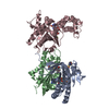
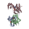
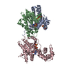
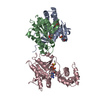
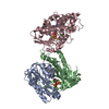
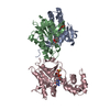
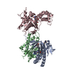
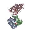
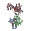
- PDB-1u0h: STRUCTURAL BASIS FOR THE INHIBITION OF MAMMALIAN ADENYLYL CYCLASE... -
+ Open data

- Basic information
| Entry | Database: PDB / ID: 1u0h | ||||||
|---|---|---|---|---|---|---|---|
| Title | STRUCTURAL BASIS FOR THE INHIBITION OF MAMMALIAN ADENYLYL CYCLASE BY MANT-GTP | ||||||
 Components Components |
| ||||||
 Keywords Keywords | LYASE / adenylyl cyclase / Gsa / MANT-GTP | ||||||
| Function / homology |  Function and homology information Function and homology informationAdenylate cyclase activating pathway / PKA activation / Hedgehog 'off' state / Adenylate cyclase inhibitory pathway / adenylate cyclase / regulation of insulin secretion involved in cellular response to glucose stimulus / sensory perception of chemical stimulus / mu-type opioid receptor binding / corticotropin-releasing hormone receptor 1 binding / adenylate cyclase activity ...Adenylate cyclase activating pathway / PKA activation / Hedgehog 'off' state / Adenylate cyclase inhibitory pathway / adenylate cyclase / regulation of insulin secretion involved in cellular response to glucose stimulus / sensory perception of chemical stimulus / mu-type opioid receptor binding / corticotropin-releasing hormone receptor 1 binding / adenylate cyclase activity / G alpha (z) signalling events / cAMP biosynthetic process / beta-2 adrenergic receptor binding / adenylate cyclase binding / D1 dopamine receptor binding / adenylate cyclase-activating adrenergic receptor signaling pathway / insulin-like growth factor receptor binding / ionotropic glutamate receptor binding / cellular response to forskolin / adenylate cyclase activator activity / G-protein beta/gamma-subunit complex binding / adenylate cyclase-modulating G protein-coupled receptor signaling pathway / manganese ion binding / adenylate cyclase-activating dopamine receptor signaling pathway / heterotrimeric G-protein complex / positive regulation of cytosolic calcium ion concentration / adenylate cyclase-activating G protein-coupled receptor signaling pathway / Hydrolases; Acting on acid anhydrides; Acting on GTP to facilitate cellular and subcellular movement / cilium / intracellular signal transduction / membrane raft / GTPase activity / dendrite / GTP binding / magnesium ion binding / protein-containing complex / ATP binding / membrane / metal ion binding / plasma membrane / cytoplasm Similarity search - Function | ||||||
| Biological species |  | ||||||
| Method |  X-RAY DIFFRACTION / SYNCHROTRON / MOLECULAR REPLACEMENT / Resolution: 2.9 Å X-RAY DIFFRACTION / SYNCHROTRON / MOLECULAR REPLACEMENT / Resolution: 2.9 Å | ||||||
 Authors Authors | Mou, T.C. / Gille, A. / Seifert, R.J. / Sprang, S.R. | ||||||
 Citation Citation | Journal: J.Biol.Chem. / Year: 2005 Title: Structural basis for the inhibition of mammalian membrane adenylyl cyclase by 2 '(3')-O-(N-Methylanthraniloyl)-guanosine 5 '-triphosphate. Authors: Mou, T.C. / Gille, A. / Fancy, D.A. / Seifert, R. / Sprang, S.R. | ||||||
| History |
|
- Structure visualization
| Structure viewer | Molecule: MolmilJmol/JSmol |
|---|
- Downloads & links
-Download
| PDBx/mmCIF format | 1u0h.cif.gz | 164.6 KB | Display | PDBx/mmCIF format |
|---|---|---|---|---|
| PDB format | pdb1u0h.ent.gz | 124.3 KB | Display | PDB format |
| PDBx/mmJSON format | 1u0h.json.gz | Tree view | PDBx/mmJSON format | |
| Others |  Other downloads Other downloads |
-Validation report
| Arichive directory | https://data.pdbj.org/pub/pdb/validation_reports/u0/1u0hftp://data.pdbj.org/pub/pdb/validation_reports/u0/1u0h | HTTPS FTP |
|---|
-Related structure data
| Related structure data |  1tl7C  1azsS C: citing same article ( S: Starting model for refinement |
|---|---|
| Similar structure data |

-Links
 PDBj
PDBj


- Assembly
| Deposited unit | 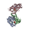
| ||||||||
|---|---|---|---|---|---|---|---|---|---|
| 1 |
| ||||||||
| Unit cell |
|
-Components
-Adenylate cyclase, type ... , 2 types, 2 molecules AB
| #1: Protein | Mass: 25526.514 Da / Num. of mol.: 1 / Fragment: C1A domain of Adenylyl Cyclase Source method: isolated from a genetically manipulated source Source: (gene. exp.)  |
|---|---|
| #2: Protein | Mass: 23717.033 Da / Num. of mol.: 1 / Fragment: C2A domain of Adenylyl Cyclase Source method: isolated from a genetically manipulated source Source: (gene. exp.) |
-Protein , 1 types, 1 molecules C
| #3: Protein | Mass: 45769.527 Da / Num. of mol.: 1 Source method: isolated from a genetically manipulated source Source: (gene. exp.) |
|---|
-Non-polymers , 6 types, 24 molecules 


| #4: Chemical |  Mass: 24.305 Da / Num. of mol.: 3 / Source method: obtained synthetically / Formula: Mg Mass: 24.305 Da / Num. of mol.: 3 / Source method: obtained synthetically / Formula: Mg#5: Chemical | ChemComp-FOK / |  Mass: 410.501 Da / Num. of mol.: 1 / Source method: obtained synthetically / Formula: C22H34O7 Mass: 410.501 Da / Num. of mol.: 1 / Source method: obtained synthetically / Formula: C22H34O7#6: Chemical | ChemComp-ONM / |  Mass: 656.328 Da / Num. of mol.: 1 / Source method: obtained synthetically / Formula: C18H23N6O15P3 Mass: 656.328 Da / Num. of mol.: 1 / Source method: obtained synthetically / Formula: C18H23N6O15P3#7: Chemical | ChemComp-CL / |  Mass: 35.453 Da / Num. of mol.: 1 / Source method: obtained synthetically / Formula: Cl Mass: 35.453 Da / Num. of mol.: 1 / Source method: obtained synthetically / Formula: Cl#8: Chemical | ChemComp-GSP / |  Mass: 539.246 Da / Num. of mol.: 1 / Source method: obtained synthetically / Formula: C10H16N5O13P3S Mass: 539.246 Da / Num. of mol.: 1 / Source method: obtained synthetically / Formula: C10H16N5O13P3S#9: Water | ChemComp-HOH / | Mass: 18.015 Da / Num. of mol.: 17 / Source method: isolated from a natural source / Formula: H2O |
|---|
-Experimental details
-Experiment
| Experiment | Method: X-RAY DIFFRACTION / Number of used crystals: 1 |
|---|
- Sample preparation
| Crystal | Density Matthews: 3.1 Å3/Da / Density % sol: 60 % |
|---|---|
| Crystal grow | Temperature: 289 K / Method: vapor diffusion, sitting drop / pH: 5.6 Details: 7.5-7.8% PEG 8000, 0.5M NACL, 0.1M PHOSPHATE BUFFER, pH 5.6, VAPOR DIFFUSION, SITTING DROP, temperature 289K |
-Data collection
| Diffraction | Mean temperature: 100 K |
|---|---|
| Diffraction source | Source: SYNCHROTRON / Site: APS  / Beamline: 19-BM / Wavelength: 1.0393 Å / Beamline: 19-BM / Wavelength: 1.0393 Å |
| Detector | Type: SBC-3 / Detector: CCD / Date: Nov 23, 2003 Details: Active area: 210 x 210 mm2 Pixel sizes: 0.079 mm (unbinned mode) and 0.159 mm (2 x 2 binned mode) Unbinned images: 3072 pixels x 3072 pixels (18.5 MB) 2 x 2 Binned images: 1536 pixels x 1536 pixels (4.6 MB) |
| Radiation | Monochromator: Si111 / Protocol: SINGLE WAVELENGTH / Monochromatic (M) / Laue (L): M / Scattering type: x-ray |
| Radiation wavelength | Wavelength: 1.0393 Å / Relative weight: 1 |
| Reflection | Resolution: 2.9→50 Å / Num. all: 24874 / Num. obs: 21911 / % possible obs: 88.1 % / Observed criterion σ(F): -2 / Observed criterion σ(I): -2 / Redundancy: 3.6 % / Biso Wilson estimate: -0.4 Å2 / Rsym value: 0.139 / Net I/σ(I): 7.8 |
| Reflection shell | Resolution: 2.91→3 Å / Redundancy: 3.1 % / Mean I/σ(I) obs: 1.9 / Num. unique all: 2245 / Rsym value: 0.37 / % possible all: 83.2 |
- Processing
| Software |
| ||||||||||||||||||||||||||||||||||||
|---|---|---|---|---|---|---|---|---|---|---|---|---|---|---|---|---|---|---|---|---|---|---|---|---|---|---|---|---|---|---|---|---|---|---|---|---|---|
| Refinement | Method to determine structure: MOLECULAR REPLACEMENT Starting model: PDB ENTRY 1AZS Resolution: 2.9→15 Å / Rfactor Rfree error: 0.01 / Data cutoff high absF: 256489.92 / Data cutoff low absF: 0 / Isotropic thermal model: RESTRAINED / Cross valid method: THROUGHOUT / σ(F): 0 / Stereochemistry target values: Engh & Huber
| ||||||||||||||||||||||||||||||||||||
| Solvent computation | Solvent model: FLAT MODEL / Bsol: 14.0723 Å2 / ksol: 0.286734 e/Å3 | ||||||||||||||||||||||||||||||||||||
| Displacement parameters | Biso mean: 54.4 Å2
| ||||||||||||||||||||||||||||||||||||
| Refine analyze |
| ||||||||||||||||||||||||||||||||||||
| Refinement step | Cycle: LAST / Resolution: 2.9→15 Å
| ||||||||||||||||||||||||||||||||||||
| Refine LS restraints |
| ||||||||||||||||||||||||||||||||||||
| LS refinement shell | Resolution: 2.9→3.08 Å / Rfactor Rfree error: 0.036 / Total num. of bins used: 6
| ||||||||||||||||||||||||||||||||||||
| Xplor file |
|
