 Movie
Movie Controller

[English] 日本語
 Yorodumi
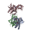
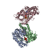
- PDB-2gvz: Crystal Structure of Complex of Gs- with The Catalytic Domains of... -
+ Open data

- Basic information
| Entry | Database: PDB / ID: 2gvz | ||||||
|---|---|---|---|---|---|---|---|
| Title | Crystal Structure of Complex of Gs- with The Catalytic Domains of Mammalian Adenylyl Cyclase: Complex with MANT-ATP and Mn | ||||||
 Components Components |
| ||||||
 Keywords Keywords | LYASE / ADENYLYL CYCLASE / MANT-ATP | ||||||
| Function / homology |  Function and homology information Function and homology informationAdenylate cyclase activating pathway / PKA activation / Hedgehog 'off' state / Adenylate cyclase inhibitory pathway / adenylate cyclase / regulation of insulin secretion involved in cellular response to glucose stimulus / sensory perception of chemical stimulus / mu-type opioid receptor binding / corticotropin-releasing hormone receptor 1 binding / adenylate cyclase activity ...Adenylate cyclase activating pathway / PKA activation / Hedgehog 'off' state / Adenylate cyclase inhibitory pathway / adenylate cyclase / regulation of insulin secretion involved in cellular response to glucose stimulus / sensory perception of chemical stimulus / mu-type opioid receptor binding / corticotropin-releasing hormone receptor 1 binding / adenylate cyclase activity / G alpha (z) signalling events / cAMP biosynthetic process / beta-2 adrenergic receptor binding / adenylate cyclase binding / D1 dopamine receptor binding / adenylate cyclase-activating adrenergic receptor signaling pathway / insulin-like growth factor receptor binding / ionotropic glutamate receptor binding / cellular response to forskolin / adenylate cyclase activator activity / G-protein beta/gamma-subunit complex binding / adenylate cyclase-modulating G protein-coupled receptor signaling pathway / manganese ion binding / adenylate cyclase-activating dopamine receptor signaling pathway / heterotrimeric G-protein complex / positive regulation of cytosolic calcium ion concentration / adenylate cyclase-activating G protein-coupled receptor signaling pathway / Hydrolases; Acting on acid anhydrides; Acting on GTP to facilitate cellular and subcellular movement / cilium / intracellular signal transduction / membrane raft / GTPase activity / dendrite / GTP binding / magnesium ion binding / protein-containing complex / ATP binding / membrane / metal ion binding / plasma membrane / cytoplasm Similarity search - Function | ||||||
| Biological species |  | ||||||
| Method |  X-RAY DIFFRACTION / SYNCHROTRON / MOLECULAR REPLACEMENT / Resolution: 3.27 Å X-RAY DIFFRACTION / SYNCHROTRON / MOLECULAR REPLACEMENT / Resolution: 3.27 Å | ||||||
 Authors Authors | Mou, T.-C. / Sprang, S.R. | ||||||
 Citation Citation | Journal: Mol.Pharmacol. / Year: 2006 Title: Broad specificity of Mammalian adenylyl cyclase for interaction with 2',3'-substituted purine- and pyrimidine nucleotide inhibitors. Authors: Mou, T.-C. / Gille, A. / Suryanarayana, S. / Richter, M. / Seifert, R. / Sprang, S.R. | ||||||
| History |
|
- Structure visualization
| Structure viewer | Molecule: MolmilJmol/JSmol |
|---|
- Downloads & links
-Download
| PDBx/mmCIF format | 2gvz.cif.gz | 164.8 KB | Display | PDBx/mmCIF format |
|---|---|---|---|---|
| PDB format | pdb2gvz.ent.gz | 124.4 KB | Display | PDB format |
| PDBx/mmJSON format | 2gvz.json.gz | Tree view | PDBx/mmJSON format | |
| Others |  Other downloads Other downloads |
-Validation report
| Arichive directory | https://data.pdbj.org/pub/pdb/validation_reports/gv/2gvzftp://data.pdbj.org/pub/pdb/validation_reports/gv/2gvz | HTTPS FTP |
|---|
-Related structure data
| Related structure data |  2gvdC  1tl7S C: citing same article ( S: Starting model for refinement |
|---|---|
| Similar structure data |


-Links
 PDBj
PDBj


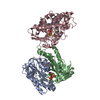
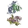
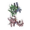
- Assembly
| Deposited unit | 
| ||||||||
|---|---|---|---|---|---|---|---|---|---|
| 1 |
| ||||||||
| Unit cell |
|
-Components
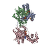
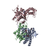
-Adenylate cyclase type ... , 2 types, 2 molecules AB
| #1: Protein | Mass: 25526.514 Da / Num. of mol.: 1 / Fragment: C1A DOMAIN, residues 440-657 Source method: isolated from a genetically manipulated source Source: (gene. exp.)  |
|---|---|
| #2: Protein | Mass: 23717.033 Da / Num. of mol.: 1 / Fragment: C2A DOMAIN, residues 870-1081 Source method: isolated from a genetically manipulated source Source: (gene. exp.) |
-Protein , 1 types, 1 molecules C
| #3: Protein | Mass: 45769.527 Da / Num. of mol.: 1 Source method: isolated from a genetically manipulated source Source: (gene. exp.) |
|---|
-Non-polymers , 5 types, 7 molecules 


| #4: Chemical |  Mass: 54.938 Da / Num. of mol.: 3 / Source method: obtained synthetically / Formula: Mn Mass: 54.938 Da / Num. of mol.: 3 / Source method: obtained synthetically / Formula: Mn#5: Chemical | ChemComp-FKP / |  Mass: 550.727 Da / Num. of mol.: 1 / Source method: obtained synthetically / Formula: C30H50N2O7 Mass: 550.727 Da / Num. of mol.: 1 / Source method: obtained synthetically / Formula: C30H50N2O7#6: Chemical | ChemComp-ONA / |  Mass: 640.328 Da / Num. of mol.: 1 / Source method: obtained synthetically / Formula: C18H23N6O14P3 Mass: 640.328 Da / Num. of mol.: 1 / Source method: obtained synthetically / Formula: C18H23N6O14P3#7: Chemical | ChemComp-CL / |  Mass: 35.453 Da / Num. of mol.: 1 / Source method: obtained synthetically / Formula: Cl Mass: 35.453 Da / Num. of mol.: 1 / Source method: obtained synthetically / Formula: Cl#8: Chemical | ChemComp-GSP / |  Mass: 539.246 Da / Num. of mol.: 1 / Source method: obtained synthetically / Formula: C10H16N5O13P3S Mass: 539.246 Da / Num. of mol.: 1 / Source method: obtained synthetically / Formula: C10H16N5O13P3S |
|---|
-Experimental details
-Experiment
| Experiment | Method: X-RAY DIFFRACTION / Number of used crystals: 2 |
|---|
- Sample preparation
| Crystal | Density Matthews: 2.82 Å3/Da / Density % sol: 56.46 % |
|---|---|
| Crystal grow | Temperature: 289 K / Method: vapor diffusion, hanging drop / pH: 5.6 Details: 7.5-7.8 %PEG8000, 0.5M NACL, 0.1M PHOSPHATE BUFFER, pH 5.6, VAPOR DIFFUSION, HANGING DROP, temperature 289K |
-Data collection
| Diffraction | Mean temperature: 100 K |
|---|---|
| Diffraction source | Source: SYNCHROTRON / Site: ALS  / Beamline: 8.2.1 / Wavelength: 1.0781 Å / Beamline: 8.2.1 / Wavelength: 1.0781 Å |
| Detector | Type: ADSC QUANTUM 210 / Detector: CCD / Date: Oct 24, 2004 / Details: KOHZU:Double Crystal Si(111) |
| Radiation | Monochromator: Si111 / Protocol: SINGLE WAVELENGTH / Monochromatic (M) / Laue (L): M / Scattering type: x-ray |
| Radiation wavelength | Wavelength: 1.0781 Å / Relative weight: 1 |
| Reflection | Resolution: 3.27→15 Å / Num. obs: 14536 |
- Processing
| Software |
| ||||||||||||||||||||||||||||||||||||
|---|---|---|---|---|---|---|---|---|---|---|---|---|---|---|---|---|---|---|---|---|---|---|---|---|---|---|---|---|---|---|---|---|---|---|---|---|---|
| Refinement | Method to determine structure: MOLECULAR REPLACEMENT Starting model: PDB ENTRY 1TL7 Resolution: 3.27→14.97 Å / Rfactor Rfree error: 0.012 / Data cutoff high absF: 166362.28 / Data cutoff low absF: 0 / Isotropic thermal model: RESTRAINED / Cross valid method: THROUGHOUT / σ(F): 0 / Stereochemistry target values: Engh & Huber
| ||||||||||||||||||||||||||||||||||||
| Solvent computation | Solvent model: FLAT MODEL / Bsol: 10 Å2 / ksol: 0.222658 e/Å3 | ||||||||||||||||||||||||||||||||||||
| Displacement parameters | Biso mean: 51.4 Å2
| ||||||||||||||||||||||||||||||||||||
| Refine analyze |
| ||||||||||||||||||||||||||||||||||||
| Refinement step | Cycle: LAST / Resolution: 3.27→14.97 Å
| ||||||||||||||||||||||||||||||||||||
| Refine LS restraints |
| ||||||||||||||||||||||||||||||||||||
| LS refinement shell | Resolution: 3.3→3.5 Å / Rfactor Rfree error: 0.044 / Total num. of bins used: 6
| ||||||||||||||||||||||||||||||||||||
| Xplor file |
|
