 Movie
Movie Controller

[English] 日本語
 Yorodumi
- PDB-3flc: Crystal structure of the His-tagged H232R mutant of glycerol kina... -
+ Open data

- Basic information
| Entry | Database: PDB / ID: 3flc | ||||||
|---|---|---|---|---|---|---|---|
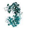
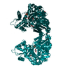
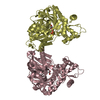
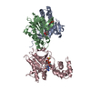
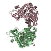
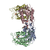
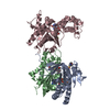
| Title | Crystal structure of the His-tagged H232R mutant of glycerol kinase from Enterococcus casseliflavus with glycerol | ||||||
 Components Components | Glycerol kinase | ||||||
 Keywords Keywords | TRANSFERASE / ATP-binding / Glycerol metabolism / Kinase / Nucleotide-binding / Phosphoprotein | ||||||
| Function / homology |  Function and homology information Function and homology informationglycerol-3-phosphate metabolic process / glycerol kinase / glycerol kinase activity / glycerol metabolic process / glycerol catabolic process / ATP binding / cytosol Similarity search - Function | ||||||
| Biological species |  Enterococcus casseliflavus (bacteria) Enterococcus casseliflavus (bacteria) | ||||||
| Method |  X-RAY DIFFRACTION / SYNCHROTRON / MOLECULAR REPLACEMENT / Resolution: 1.85 Å X-RAY DIFFRACTION / SYNCHROTRON / MOLECULAR REPLACEMENT / Resolution: 1.85 Å | ||||||
 Authors Authors | Briozzo, P. | ||||||
 Citation Citation | Journal: Biochemistry / Year: 2008 Title: Structural Characterizations of Glycerol Kinase: Unraveling Phosphorylation-Induced Long-Range Activation Authors: Yeh, J.I. / Kettering, R. / Saxl, R. / Bourand, A. / Darbon, E. / Joly, N. / Briozzo, P. / Deutscher, J. | ||||||
| History |
|
- Structure visualization
| Structure viewer | Molecule: MolmilJmol/JSmol |
|---|
- Downloads & links
-Download
| PDBx/mmCIF format | 3flc.cif.gz | 215.6 KB | Display | PDBx/mmCIF format |
|---|---|---|---|---|
| PDB format | pdb3flc.ent.gz | 171.3 KB | Display | PDB format |
| PDBx/mmJSON format | 3flc.json.gz | Tree view | PDBx/mmJSON format | |
| Others |  Other downloads Other downloads |
-Validation report
| Arichive directory | https://data.pdbj.org/pub/pdb/validation_reports/fl/3flcftp://data.pdbj.org/pub/pdb/validation_reports/fl/3flc | HTTPS FTP |
|---|
-Related structure data
| Related structure data |  3d7eC  3h3nC  3h3oC 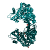 3h45C  3h46C 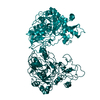 1r59S S: Starting model for refinement C: citing same article ( |
|---|---|
| Similar structure data |


-Links
 PDBj
PDBj- Assembly
| Deposited unit | 
| ||||||||
|---|---|---|---|---|---|---|---|---|---|
| 1 |
| ||||||||
| Unit cell |
|
-Components
| #1: Protein | Mass: 57237.070 Da / Num. of mol.: 2 / Mutation: H232R Source method: isolated from a genetically manipulated source Source: (gene. exp.) Enterococcus casseliflavus (bacteria) / Gene: glpK / Plasmid: pOXO4 / Production host: #2: Chemical |   Mass: 92.094 Da / Num. of mol.: 2 / Source method: obtained synthetically / Formula: C3H8O3 Mass: 92.094 Da / Num. of mol.: 2 / Source method: obtained synthetically / Formula: C3H8O3#3: Chemical |   Mass: 96.063 Da / Num. of mol.: 2 / Source method: obtained synthetically / Formula: SO4 Mass: 96.063 Da / Num. of mol.: 2 / Source method: obtained synthetically / Formula: SO4#4: Water | ChemComp-HOH / |  Mass: 18.015 Da / Num. of mol.: 708 / Source method: isolated from a natural source / Formula: H2O Mass: 18.015 Da / Num. of mol.: 708 / Source method: isolated from a natural source / Formula: H2O |
|---|
-Experimental details
-Experiment
| Experiment | Method: X-RAY DIFFRACTION / Number of used crystals: 1 |
|---|
- Sample preparation
| Crystal | Density Matthews: 2.25 Å3/Da / Density % sol: 45.25 % |
|---|---|
| Crystal grow | Temperature: 293 K / Method: vapor diffusion, hanging drop / pH: 7.5 Details: 2.0M ammonium sulfate, 5%(v/v) isopropanol, pH 7.5, VAPOR DIFFUSION, HANGING DROP, temperature 293K |
-Data collection
| Diffraction | Mean temperature: 100 K |
|---|---|
| Diffraction source | Source: SYNCHROTRON / Site: ESRF  / Beamline: ID14-3 / Wavelength: 0.931 Å / Beamline: ID14-3 / Wavelength: 0.931 Å |
| Detector | Detector: CCD / Date: Mar 17, 2005 |
| Radiation | Protocol: SINGLE WAVELENGTH / Monochromatic (M) / Laue (L): M / Scattering type: x-ray |
| Radiation wavelength | Wavelength: 0.931 Å / Relative weight: 1 |
| Reflection | Resolution: 1.85→30 Å / Num. obs: 87047 / % possible obs: 97.8 % / Observed criterion σ(I): 2 / Redundancy: 18.25 % / Rsym value: 0.073 / Net I/σ(I): 16.2 |
| Reflection shell | Resolution: 1.85→1.92 Å / Mean I/σ(I) obs: 2.15 / Rsym value: 0.598 / % possible all: 97.8 |
- Processing
| Software |
| ||||||||||||||||||||
|---|---|---|---|---|---|---|---|---|---|---|---|---|---|---|---|---|---|---|---|---|---|
| Refinement | Method to determine structure: MOLECULAR REPLACEMENT Starting model: PDB ENTRY 1R59, molecule O Resolution: 1.85→30 Å / σ(F): 2 / Stereochemistry target values: CNS
| ||||||||||||||||||||
| Refinement step | Cycle: LAST / Resolution: 1.85→30 Å
| ||||||||||||||||||||
| Refine LS restraints |
|
