Stage V sporulation protein AD / Stage V sporulation AD / Stage V sporulation AD superfamily / Stage V sporulation protein AD (SpoVAD) / Peroxisomal Thiolase; Chain A, domain 1 / Thiolase-like / 3-Layer(aba) Sandwich / Alpha Beta Similarity search - Domain/homology
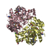
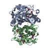
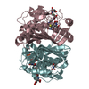
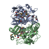
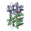
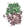
A: Stage V sporulation protein AD (SpoVAD) B: Stage V sporulation protein AD (SpoVAD) C: Stage V sporulation protein AD (SpoVAD) D: Stage V sporulation protein AD (SpoVAD)
Resolution: 2→2.07 Å / Redundancy: 2.7 % / Rmerge(I) obs: 0.201 / Mean I/σ(I) obs: 4.53 / Num. unique all: 14586 / % possible all: 84.2
-
Processing
Software
Name
Version
Classification
PHENIX
(phenix.refine: 1.5_2)
refinement
SHELXDE
phasing
SOLVE
phasing
CNS
refinement
HKL-2000
datacollection
DENZO
datareduction
SCALEPACK
datascaling
Refinement
Method to determine structure: SAD / Resolution: 1.993→38.939 Å / SU ML: 0.23 / Cross valid method: THROUGHOUT / σ(F): 1.34 / Stereochemistry target values: ML
Rfactor
Num. reflection
% reflection
Selection details
Rfree
0.2109
3683
5.03 %
RANDOM
Rwork
0.1681
-
-
-
obs
0.1703
73163
98.95 %
-
Solvent computation
Shrinkage radii: 0.9 Å / VDW probe radii: 1.11 Å / Solvent model: FLAT BULK SOLVENT MODEL / Bsol: 28.18 Å2 / ksol: 0.32 e/Å3
Refinement step
Cycle: LAST / Resolution: 1.993→38.939 Å
Protein
Nucleic acid
Ligand
Solvent
Total
Num. atoms
9375
0
0
587
9962
Refine LS restraints
Refine-ID
Type
Dev ideal
Number
X-RAY DIFFRACTION
f_bond_d
0.008
9524
X-RAY DIFFRACTION
f_angle_d
1.378
12881
X-RAY DIFFRACTION
f_dihedral_angle_d
17.565
3438
X-RAY DIFFRACTION
f_chiral_restr
0.1
1508
X-RAY DIFFRACTION
f_plane_restr
0.005
1668
LS refinement shell
Resolution (Å)
Rfactor Rfree
Num. reflection Rfree
Rfactor Rwork
Num. reflection Rwork
Refine-ID
% reflection obs (%)
1.9928-2.019
0.2708
132
0.1929
2315
X-RAY DIFFRACTION
87
2.019-2.0466
0.2262
143
0.172
2659
X-RAY DIFFRACTION
99
2.0466-2.0759
0.2349
128
0.1632
2698
X-RAY DIFFRACTION
99
2.0759-2.1069
0.23
122
0.1697
2689
X-RAY DIFFRACTION
99
2.1069-2.1398
0.2428
129
0.1633
2647
X-RAY DIFFRACTION
99
2.1398-2.1749
0.2085
133
0.1657
2688
X-RAY DIFFRACTION
99
2.1749-2.2124
0.224
157
0.1692
2622
X-RAY DIFFRACTION
99
2.2124-2.2526
0.2557
126
0.1707
2697
X-RAY DIFFRACTION
99
2.2526-2.2959
0.2331
138
0.1834
2643
X-RAY DIFFRACTION
99
2.2959-2.3428
0.2176
149
0.165
2659
X-RAY DIFFRACTION
99
2.3428-2.3937
0.2347
145
0.1751
2671
X-RAY DIFFRACTION
99
2.3937-2.4494
0.2187
128
0.1788
2703
X-RAY DIFFRACTION
100
2.4494-2.5106
0.256
158
0.18
2696
X-RAY DIFFRACTION
100
2.5106-2.5785
0.245
145
0.1705
2658
X-RAY DIFFRACTION
100
2.5785-2.6543
0.2093
137
0.1686
2702
X-RAY DIFFRACTION
100
2.6543-2.74
0.2198
146
0.1699
2669
X-RAY DIFFRACTION
100
2.74-2.8379
0.2301
146
0.1789
2727
X-RAY DIFFRACTION
100
2.8379-2.9515
0.223
150
0.176
2707
X-RAY DIFFRACTION
100
2.9515-3.0858
0.2275
122
0.1821
2725
X-RAY DIFFRACTION
100
3.0858-3.2484
0.2313
148
0.1699
2693
X-RAY DIFFRACTION
100
3.2484-3.4518
0.1902
148
0.1637
2674
X-RAY DIFFRACTION
100
3.4518-3.7181
0.1883
165
0.1586
2684
X-RAY DIFFRACTION
100
3.7181-4.0919
0.1775
161
0.149
2696
X-RAY DIFFRACTION
100
4.0919-4.6832
0.1614
148
0.1305
2689
X-RAY DIFFRACTION
99
4.6832-5.897
0.1631
136
0.1532
2746
X-RAY DIFFRACTION
100
5.897-38.9469
0.2094
143
0.178
2723
X-RAY DIFFRACTION
98
Refinement TLS params.
Refine-ID: X-RAY DIFFRACTION
ID
Method
L11 (°2)
L12 (°2)
L13 (°2)
L22 (°2)
L23 (°2)
L33 (°2)
S11 (Å °)
S12 (Å °)
S13 (Å °)
S21 (Å °)
S22 (Å °)
S23 (Å °)
S31 (Å °)
S32 (Å °)
S33 (Å °)
T11 (Å2)
T12 (Å2)
T13 (Å2)
T22 (Å2)
T23 (Å2)
T33 (Å2)
Origin x (Å)
Origin y (Å)
Origin z (Å)
1
refined
0.0828
0.0445
0.0183
0.0149
0.0464
-0.0161
-0.1229
-0.2322
-0.0276
-0.0149
-0.021
-0.0575
0.0645
-0.0938
-0
-0.1962
-0.2865
-0.0772
-0.3634
-0.1163
-0.0212
20.1983
44.5389
40.2786
2
0.1369
0.0642
-0.0077
0.0059
0.0042
0.0191
-0.1212
0.0034
-0.0237
0.0785
0.0642
0.0906
0.0352
0.1246
0
-0.1061
0.0785
-0.0686
0.059
0.0407
-0.0692
3
0.1148
0.0069
0.0384
-0.0032
0.0077
0.0528
-0.0283
0.0407
-0.0554
0.0211
-0.007
0.0087
0.0321
-0.0654
-0
0.0522
-0.0043
0.0234
-0.0242
0.0089
0.0348
4
0.1341
0.0878
0.0076
0.0308
0.0218
0.0398
-0.0304
-0.0501
0.0157
-0.0049
-0.0114
-0.0258
0.0158
0.0177
-0
0.0599
-0.0016
-0.0062
0.0759
-0.0162
0.0732
Refinement TLS group
Selection details: chain D
+
About Yorodumi
-
News
-
Feb 9, 2022. New format data for meta-information of EMDB entries
New format data for meta-information of EMDB entries
Version 3 of the EMDB header file is now the official format.
The previous official version 1.9 will be removed from the archive.
In the structure databanks used in Yorodumi, some data are registered as the other names, "COVID-19 virus" and "2019-nCoV". Here are the details of the virus and the list of structure data.
Jan 31, 2019. EMDB accession codes are about to change! (news from PDBe EMDB page)
EMDB accession codes are about to change! (news from PDBe EMDB page)
The allocation of 4 digits for EMDB accession codes will soon come to an end. Whilst these codes will remain in use, new EMDB accession codes will include an additional digit and will expand incrementally as the available range of codes is exhausted. The current 4-digit format prefixed with “EMD-” (i.e. EMD-XXXX) will advance to a 5-digit format (i.e. EMD-XXXXX), and so on. It is currently estimated that the 4-digit codes will be depleted around Spring 2019, at which point the 5-digit format will come into force.
The EM Navigator/Yorodumi systems omit the EMD- prefix.
Related info.:Q: What is EMD? / ID/Accession-code notation in Yorodumi/EM Navigator
Yorodumi is a browser for structure data from EMDB, PDB, SASBDB, etc.
This page is also the successor to EM Navigator detail page, and also detail information page/front-end page for Omokage search.
The word "yorodu" (or yorozu) is an old Japanese word meaning "ten thousand". "mi" (miru) is to see.
Related info.:EMDB / PDB / SASBDB / Comparison of 3 databanks / Yorodumi Search / Aug 31, 2016. New EM Navigator & Yorodumi / Yorodumi Papers / Jmol/JSmol / Function and homology information / Changes in new EM Navigator and Yorodumi
 Movie
Movie Controller

 Yorodumi
 Open data
 Basic information
 Components
 Keywords
 Function and homology information
 X-RAY DIFFRACTION /
  Authors
 Citation
 Structure visualization
 Downloads & links
 Other downloads

 PDBj
PDBj

 Assembly
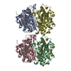

 Mass: 18.015 Da / Num. of mol.: 587 / Source method: isolated from a natural source / Formula: H2O
 Sample preparation
 / Beamline: X4C / Wavelength: 0.97916 Å
 Processing