 Movie
Movie Controller

[English] 日本語
 Yorodumi
- PDB-6ulo: Structure of an N-terminally truncated uncharacterized protein fr... -
+ Open data

- Basic information
| Entry | Database: PDB / ID: 6ulo | ||||||
|---|---|---|---|---|---|---|---|
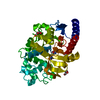
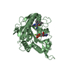
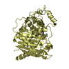
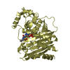
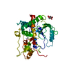
| Title | Structure of an N-terminally truncated uncharacterized protein from Leptospira interrogans serogroup Icterohaemorrhagiae serovar Copenhageni (strain Fiocruz L1-130) | ||||||
 Components Components | Uncharacterized protein | ||||||
 Keywords Keywords | UNKNOWN FUNCTION / Structural Genomics / Seattle Structural Genomics Center for Infectious Disease / SSGCID | ||||||
| Function / homology | membrane / Lipoprotein Function and homology information Function and homology information | ||||||
| Biological species |  Leptospira interrogans serogroup Icterohaemorrhagiae serovar copenhageni (bacteria) Leptospira interrogans serogroup Icterohaemorrhagiae serovar copenhageni (bacteria) | ||||||
| Method |  X-RAY DIFFRACTION / SYNCHROTRON / SAD / Resolution: 1.3 Å X-RAY DIFFRACTION / SYNCHROTRON / SAD / Resolution: 1.3 Å | ||||||
 Authors Authors | Seattle Structural Genomics Center for Infectious Disease (SSGCID) | ||||||
 Citation Citation | Journal: to be published Title: Structure of an N-terminally truncated uncharacterized protein from Leptospira interrogans serogroup Icterohaemorrhagiae serovar Copenhageni (strain Fiocruz L1-130) Authors: Abendroth, J. / Mayclin, S.J. / Lorimer, D.D. / Horanyi, P.S. / Edwards, T.E. | ||||||
| History |
|
- Structure visualization
| Structure viewer | Molecule: MolmilJmol/JSmol |
|---|
- Downloads & links
-Download
| PDBx/mmCIF format | 6ulo.cif.gz | 343.3 KB | Display | PDBx/mmCIF format |
|---|---|---|---|---|
| PDB format | pdb6ulo.ent.gz | 229.8 KB | Display | PDB format |
| PDBx/mmJSON format | 6ulo.json.gz | Tree view | PDBx/mmJSON format | |
| Others |  Other downloads Other downloads |
-Validation report
| Arichive directory | https://data.pdbj.org/pub/pdb/validation_reports/ul/6uloftp://data.pdbj.org/pub/pdb/validation_reports/ul/6ulo | HTTPS FTP |
|---|
-Related structure data
| Similar structure data | |
|---|---|
| Other databases |


-Links
 PDBj
PDBj- Assembly
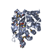
| Deposited unit | 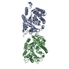
| ||||||||||||
|---|---|---|---|---|---|---|---|---|---|---|---|---|---|
| 1 | 
| ||||||||||||
| 2 | 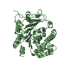
| ||||||||||||
| Unit cell |
|
-Components
| #1: Protein | Mass: 38038.465 Da / Num. of mol.: 2 Source method: isolated from a genetically manipulated source Source: (gene. exp.) Leptospira interrogans serogroup Icterohaemorrhagiae serovar copenhageni (strain Fiocruz L1-130) (bacteria)Strain: Fiocruz L1-130 / Gene: LIC_10985 / Plasmid: LpinA.19565.a.B2 Production host: Strain (production host): BL21(DE3) / References: UniProt: Q72TN2 #2: Chemical |   Mass: 35.453 Da / Num. of mol.: 2 / Source method: isolated from a natural source / Formula: Cl Mass: 35.453 Da / Num. of mol.: 2 / Source method: isolated from a natural source / Formula: Cl#3: Water | ChemComp-HOH / |  Mass: 18.015 Da / Num. of mol.: 1021 / Source method: isolated from a natural source / Formula: H2O Mass: 18.015 Da / Num. of mol.: 1021 / Source method: isolated from a natural source / Formula: H2OHas ligand of interest | N | |
|---|
-Experimental details
-Experiment
| Experiment | Method: X-RAY DIFFRACTION / Number of used crystals: 1 |
|---|
- Sample preparation
| Crystal | Density Matthews: 2.26 Å3/Da / Density % sol: 45.5 % |
|---|---|
| Crystal grow | Temperature: 290 K / Method: vapor diffusion, sitting drop / pH: 9.5 Details: Molecular Dimensions Morpheus screen, condition H5: 10% w/v PEG 20 000, 20% v/v PEG MME 550: 20mM of each amino acid: sodium l-glutamate, DL-alanine, glycine, DL-lysine HCl, DL-serine: 100mM ...Details: Molecular Dimensions Morpheus screen, condition H5: 10% w/v PEG 20 000, 20% v/v PEG MME 550: 20mM of each amino acid: sodium l-glutamate, DL-alanine, glycine, DL-lysine HCl, DL-serine: 100mM MOPS/HEPES-Na pH 7.5: LpinA.195645.a.B2.PW386228 at 23mg/ml. Cryo: direct: tray 310523 h5: puck PAS1-8. For phasing a crystal from the same tray, well C1 (10% w/v PEG 20 000, 20% v/v PEG MME 550: 30mM of each sodium nitrate, disodium hydrogen phosphate, ammonium sulfate: 100mM MES/imidazole pH 6.5) was incubated for 20sec in a mixture of 10% 5M sodium iodide in ethylene glycol (500mM final NaI concentration) and 90% reservoir solution. Diffraction data were collected in-house.Tray 310523 c1: puck LOF7-9 |
-Data collection
| Diffraction |
| ||||||||||||||||||||||||||||||||||||||||||||||||||||||||||||||||||||||||||||||||||||||||||||||||||||||||||||||||||||||||||||||||||||||||||||||||||||||||||||||||||||||||
|---|---|---|---|---|---|---|---|---|---|---|---|---|---|---|---|---|---|---|---|---|---|---|---|---|---|---|---|---|---|---|---|---|---|---|---|---|---|---|---|---|---|---|---|---|---|---|---|---|---|---|---|---|---|---|---|---|---|---|---|---|---|---|---|---|---|---|---|---|---|---|---|---|---|---|---|---|---|---|---|---|---|---|---|---|---|---|---|---|---|---|---|---|---|---|---|---|---|---|---|---|---|---|---|---|---|---|---|---|---|---|---|---|---|---|---|---|---|---|---|---|---|---|---|---|---|---|---|---|---|---|---|---|---|---|---|---|---|---|---|---|---|---|---|---|---|---|---|---|---|---|---|---|---|---|---|---|---|---|---|---|---|---|---|---|---|---|---|---|---|
| Diffraction source |
| ||||||||||||||||||||||||||||||||||||||||||||||||||||||||||||||||||||||||||||||||||||||||||||||||||||||||||||||||||||||||||||||||||||||||||||||||||||||||||||||||||||||||
| Detector |
| ||||||||||||||||||||||||||||||||||||||||||||||||||||||||||||||||||||||||||||||||||||||||||||||||||||||||||||||||||||||||||||||||||||||||||||||||||||||||||||||||||||||||
| Radiation |
| ||||||||||||||||||||||||||||||||||||||||||||||||||||||||||||||||||||||||||||||||||||||||||||||||||||||||||||||||||||||||||||||||||||||||||||||||||||||||||||||||||||||||
| Radiation wavelength |
| ||||||||||||||||||||||||||||||||||||||||||||||||||||||||||||||||||||||||||||||||||||||||||||||||||||||||||||||||||||||||||||||||||||||||||||||||||||||||||||||||||||||||
| Reflection | Resolution: 1.3→50 Å / Num. obs: 155840 / % possible obs: 95.3 % / Redundancy: 3.927 % / Biso Wilson estimate: 19.615 Å2 / CC1/2: 1 / Rmerge(I) obs: 0.036 / Rrim(I) all: 0.041 / Χ2: 1.052 / Net I/σ(I): 19.58 | ||||||||||||||||||||||||||||||||||||||||||||||||||||||||||||||||||||||||||||||||||||||||||||||||||||||||||||||||||||||||||||||||||||||||||||||||||||||||||||||||||||||||
| Reflection shell | Diffraction-ID: 1
|

- Processing
| Software |
| |||||||||||||||||||||||||||||||||||||||||||||||||||||||||||||||||||||||||||||||||||||||||||||||||||||||||
|---|---|---|---|---|---|---|---|---|---|---|---|---|---|---|---|---|---|---|---|---|---|---|---|---|---|---|---|---|---|---|---|---|---|---|---|---|---|---|---|---|---|---|---|---|---|---|---|---|---|---|---|---|---|---|---|---|---|---|---|---|---|---|---|---|---|---|---|---|---|---|---|---|---|---|---|---|---|---|---|---|---|---|---|---|---|---|---|---|---|---|---|---|---|---|---|---|---|---|---|---|---|---|---|---|---|---|
| Refinement | Method to determine structure: SAD / Resolution: 1.3→32.24 Å / SU ML: 0.1214 / Cross valid method: FREE R-VALUE / σ(F): 1.98 / Phase error: 17.6941 Stereochemistry target values: GeoStd + Monomer Library + CDL v1.2
| |||||||||||||||||||||||||||||||||||||||||||||||||||||||||||||||||||||||||||||||||||||||||||||||||||||||||
| Solvent computation | Shrinkage radii: 0.9 Å / VDW probe radii: 1.11 Å / Solvent model: FLAT BULK SOLVENT MODEL | |||||||||||||||||||||||||||||||||||||||||||||||||||||||||||||||||||||||||||||||||||||||||||||||||||||||||
| Displacement parameters | Biso mean: 19.46 Å2 | |||||||||||||||||||||||||||||||||||||||||||||||||||||||||||||||||||||||||||||||||||||||||||||||||||||||||
| Refinement step | Cycle: LAST / Resolution: 1.3→32.24 Å
| |||||||||||||||||||||||||||||||||||||||||||||||||||||||||||||||||||||||||||||||||||||||||||||||||||||||||
| Refine LS restraints |
| |||||||||||||||||||||||||||||||||||||||||||||||||||||||||||||||||||||||||||||||||||||||||||||||||||||||||
| LS refinement shell |
|
