 Movie
Movie Controller

[English] 日本語
 Yorodumi
- PDB-4kkv: Crystal structure of candida glabrata FMN adenylyltransferase D18... -
+ Open data

- Basic information
| Entry | Database: PDB / ID: 4kkv | ||||||
|---|---|---|---|---|---|---|---|
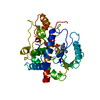
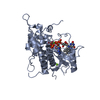
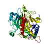
| Title | Crystal structure of candida glabrata FMN adenylyltransferase D181A Mutant | ||||||
 Components Components | Similar to uniprot|P38913 Saccharomyces cerevisiae YDL045c FAD synthetase | ||||||
 Keywords Keywords | TRANSFERASE / alpha/beta protein / Rossmann-like fold / FAD biosynthesis | ||||||
| Function / homology |  Function and homology information Function and homology informationFAD synthase / FMN adenylyltransferase activity / FAD biosynthetic process / magnesium ion binding / ATP binding / cytoplasm Similarity search - Function | ||||||
| Biological species |  Candida glabrata (fungus) Candida glabrata (fungus) | ||||||
| Method |  X-RAY DIFFRACTION / MOLECULAR REPLACEMENT / Resolution: 1.74 Å X-RAY DIFFRACTION / MOLECULAR REPLACEMENT / Resolution: 1.74 Å | ||||||
 Authors Authors | Huerta, C. / Zhang, H. | ||||||
 Citation Citation | Journal: Biochemistry / Year: 2013 Title: The "Super Mutant" of Yeast FMN Adenylyltransferase Enhances the Enzyme Turnover Rate by Attenuating Product Inhibition. Authors: Huerta, C. / Grishin, N.V. / Zhang, H. | ||||||
| History |
|
- Structure visualization
| Structure viewer | Molecule: MolmilJmol/JSmol |
|---|
- Downloads & links
-Download
| PDBx/mmCIF format | 4kkv.cif.gz | 85.3 KB | Display | PDBx/mmCIF format |
|---|---|---|---|---|
| PDB format | pdb4kkv.ent.gz | 63.6 KB | Display | PDB format |
| PDBx/mmJSON format | 4kkv.json.gz | Tree view | PDBx/mmJSON format | |
| Others |  Other downloads Other downloads |
-Validation report
| Arichive directory | https://data.pdbj.org/pub/pdb/validation_reports/kk/4kkvftp://data.pdbj.org/pub/pdb/validation_reports/kk/4kkv | HTTPS FTP |
|---|
-Related structure data
| Related structure data | 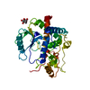 3fwkS S: Starting model for refinement |
|---|---|
| Similar structure data |


-Links
 PDBj
PDBj


- Assembly
| Deposited unit | 
| ||||||||
|---|---|---|---|---|---|---|---|---|---|
| 1 |
| ||||||||
| Unit cell |
| ||||||||
| Components on special symmetry positions |
|
-Components
| #1: Protein | Mass: 35941.715 Da / Num. of mol.: 1 / Mutation: D181A Source method: isolated from a genetically manipulated source Source: (gene. exp.) Candida glabrata (fungus) / Strain: NCYC / Gene: CAGL0K01397g, FAD1 / Plasmid: pHIS parallel / Production host:  |
|---|---|
| #2: Sugar | ChemComp-BGC /   Type: D-saccharide, beta linking / Mass: 180.156 Da / Num. of mol.: 1 Type: D-saccharide, beta linking / Mass: 180.156 Da / Num. of mol.: 1Source method: isolated from a genetically manipulated source Formula: C6H12O6 |
| #3: Chemical | ChemComp-CL /   Mass: 35.453 Da / Num. of mol.: 1 / Source method: obtained synthetically / Formula: Cl Mass: 35.453 Da / Num. of mol.: 1 / Source method: obtained synthetically / Formula: Cl |
| #4: Water | ChemComp-HOH /  Mass: 18.015 Da / Num. of mol.: 332 / Source method: isolated from a natural source / Formula: H2O Mass: 18.015 Da / Num. of mol.: 332 / Source method: isolated from a natural source / Formula: H2O |
-Experimental details
-Experiment
| Experiment | Method: X-RAY DIFFRACTION / Number of used crystals: 1 |
|---|
- Sample preparation
| Crystal | Density Matthews: 2.02 Å3/Da / Density % sol: 39.17 % |
|---|---|
| Crystal grow | Temperature: 289 K / Method: vapor diffusion, hanging drop / pH: 5 Details: 0.1 M Sodium acetate, pH 5.0, 8% w/v PEG 4000, VAPOR DIFFUSION, HANGING DROP, temperature 289K |
-Data collection
| Diffraction | Mean temperature: 93 K |
|---|---|
| Diffraction source | Source: ROTATING ANODE / Type: RIGAKU FR-E SUPERBRIGHT / Wavelength: 1.54178 Å |
| Detector | Type: RIGAKU RAXIS IV / Detector: IMAGE PLATE / Date: Jul 15, 2010 / Details: Mirrors |
| Radiation | Monochromator: Mirrors / Protocol: SINGLE WAVELENGTH / Monochromatic (M) / Laue (L): M / Scattering type: x-ray |
| Radiation wavelength | Wavelength: 1.54178 Å / Relative weight: 1 |
| Reflection | Resolution: 1.74→50 Å / Num. all: 30336 / Num. obs: 30119 / % possible obs: 99.3 % / Observed criterion σ(F): 0 / Observed criterion σ(I): 3 / Redundancy: 5.5 % / Biso Wilson estimate: 17.85 Å2 / Rmerge(I) obs: 0.053 / Net I/σ(I): 21.6 |
| Reflection shell | Resolution: 1.74→1.77 Å / Redundancy: 3.5 % / Rmerge(I) obs: 0.497 / Mean I/σ(I) obs: 1.26 / Num. unique all: 1410 / % possible all: 95.4 |
- Processing
| Software |
| ||||||||||||||||||||||||||||||||||||||||||||||||||||||||||||
|---|---|---|---|---|---|---|---|---|---|---|---|---|---|---|---|---|---|---|---|---|---|---|---|---|---|---|---|---|---|---|---|---|---|---|---|---|---|---|---|---|---|---|---|---|---|---|---|---|---|---|---|---|---|---|---|---|---|---|---|---|---|
| Refinement | Method to determine structure: MOLECULAR REPLACEMENT Starting model: PDB Entry 3FWK Resolution: 1.74→24.4 Å / Cor.coef. Fo:Fc: 0.964 / Cor.coef. Fo:Fc free: 0.951 / SU B: 2.346 / SU ML: 0.077 / Isotropic thermal model: Isotropic / Cross valid method: THROUGHOUT / ESU R: 0.128 / ESU R Free: 0.115 / Stereochemistry target values: MAXIMUM LIKELIHOOD / Details: HYDROGENS HAVE BEEN ADDED IN THE RIDING POSITIONS
| ||||||||||||||||||||||||||||||||||||||||||||||||||||||||||||
| Solvent computation | Ion probe radii: 0.8 Å / Shrinkage radii: 0.8 Å / VDW probe radii: 1.2 Å / Solvent model: MASK | ||||||||||||||||||||||||||||||||||||||||||||||||||||||||||||
| Displacement parameters | Biso mean: 22.121 Å2
| ||||||||||||||||||||||||||||||||||||||||||||||||||||||||||||
| Refinement step | Cycle: LAST / Resolution: 1.74→24.4 Å
| ||||||||||||||||||||||||||||||||||||||||||||||||||||||||||||
| Refine LS restraints |
| ||||||||||||||||||||||||||||||||||||||||||||||||||||||||||||
| LS refinement shell | Resolution: 1.74→1.785 Å / Total num. of bins used: 20
|
