 Movie
Movie Controller

[English] 日本語
 Yorodumi
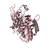
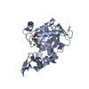
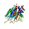
- PDB-3g6k: Crystal Structure of Candida glabrata FMN Adenylyltransferase in ... -
+ Open data

- Basic information
| Entry | Database: PDB / ID: 3g6k | ||||||
|---|---|---|---|---|---|---|---|
| Title | Crystal Structure of Candida glabrata FMN Adenylyltransferase in complex with FAD and Inorganic Pyrophosphate | ||||||
 Components Components | FMN adenylyltransferase | ||||||
 Keywords Keywords | TRANSFERASE / FAD binding / FAD biosynthesis / alpha/beta protein / Rossmann-like fold / extended loop region | ||||||
| Function / homology |  Function and homology information Function and homology informationFAD synthase / FMN adenylyltransferase activity / FAD biosynthetic process / magnesium ion binding / ATP binding / cytoplasm Similarity search - Function | ||||||
| Biological species |  Candida glabrata (fungus) Candida glabrata (fungus) | ||||||
| Method |  X-RAY DIFFRACTION / SYNCHROTRON / MOLECULAR REPLACEMENT / Resolution: 1.35 Å X-RAY DIFFRACTION / SYNCHROTRON / MOLECULAR REPLACEMENT / Resolution: 1.35 Å | ||||||
 Authors Authors | Huerta, C. / Machius, M. / Zhang, H. | ||||||
 Citation Citation | Journal: J.Mol.Biol. / Year: 2009 Title: Structure and mechanism of a eukaryotic FMN adenylyltransferase. Authors: Huerta, C. / Borek, D. / Machius, M. / Grishin, N.V. / Zhang, H. | ||||||
| History |
|
- Structure visualization
| Structure viewer | Molecule: MolmilJmol/JSmol |
|---|
- Downloads & links
-Download
| PDBx/mmCIF format | 3g6k.cif.gz | 683.9 KB | Display | PDBx/mmCIF format |
|---|---|---|---|---|
| PDB format | pdb3g6k.ent.gz | 564.6 KB | Display | PDB format |
| PDBx/mmJSON format | 3g6k.json.gz | Tree view | PDBx/mmJSON format | |
| Others |  Other downloads Other downloads |
-Validation report
| Arichive directory | https://data.pdbj.org/pub/pdb/validation_reports/g6/3g6kftp://data.pdbj.org/pub/pdb/validation_reports/g6/3g6k | HTTPS FTP |
|---|
-Related structure data
| Related structure data |  3fwkSC  3g59C  3g5aC S: Starting model for refinement C: citing same article ( |
|---|---|
| Similar structure data |


-Links
 PDBj
PDBj


- Assembly
| Deposited unit | 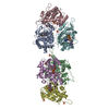
| |||||||||
|---|---|---|---|---|---|---|---|---|---|---|
| 1 | 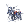
| |||||||||
| 2 | 
| |||||||||
| 3 | 
| |||||||||
| 4 | 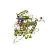
| |||||||||
| 5 | 
| |||||||||
| 6 | 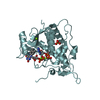
| |||||||||
| Unit cell |
| |||||||||
| Components on special symmetry positions |
|
-Components
-Protein / Sugars , 2 types, 7 molecules ABCDEF
| #1: Protein | Mass: 35985.723 Da / Num. of mol.: 6 Source method: isolated from a genetically manipulated source Source: (gene. exp.) Candida glabrata (fungus) / Strain: NCYC / Gene: CAGL0K01397g, FAD1 / Plasmid: pHIS parallel / Production host:  #6: Sugar | ChemComp-BGC / |  Type: D-saccharide, beta linking / Mass: 180.156 Da / Num. of mol.: 1 Type: D-saccharide, beta linking / Mass: 180.156 Da / Num. of mol.: 1Source method: isolated from a genetically manipulated source Formula: C6H12O6 |
|---|
-Non-polymers , 5 types, 1899 molecules 


| #2: Chemical | ChemComp-POP /  Mass: 175.959 Da / Num. of mol.: 6 / Source method: obtained synthetically / Formula: H2O7P2 Mass: 175.959 Da / Num. of mol.: 6 / Source method: obtained synthetically / Formula: H2O7P2#3: Chemical | ChemComp-FAD /  Mass: 785.550 Da / Num. of mol.: 6 / Source method: obtained synthetically / Formula: C27H33N9O15P2 / Comment: FAD*YM Mass: 785.550 Da / Num. of mol.: 6 / Source method: obtained synthetically / Formula: C27H33N9O15P2 / Comment: FAD*YM#4: Chemical | ChemComp-MG /  Mass: 24.305 Da / Num. of mol.: 13 / Source method: obtained synthetically / Formula: Mg Mass: 24.305 Da / Num. of mol.: 13 / Source method: obtained synthetically / Formula: Mg#5: Chemical | ChemComp-SO4 /  Mass: 96.063 Da / Num. of mol.: 4 / Source method: obtained synthetically / Formula: SO4 Mass: 96.063 Da / Num. of mol.: 4 / Source method: obtained synthetically / Formula: SO4#7: Water | ChemComp-HOH / | Mass: 18.015 Da / Num. of mol.: 1870 / Source method: isolated from a natural source / Formula: H2O |
|---|
-Experimental details
-Experiment
| Experiment | Method: X-RAY DIFFRACTION / Number of used crystals: 1 |
|---|
- Sample preparation
| Crystal | Density Matthews: 2.05 Å3/Da / Density % sol: 39.88 % |
|---|---|
| Crystal grow | Temperature: 293 K / Method: vapor diffusion, hanging drop / pH: 4.8 Details: 22% (w/v) PEG MME 2000, 0.2 M Magnesium sulfate, 0.1 M Sodium acetate, pH 4.8, Temperature 293K, VAPOR DIFFUSION, HANGING DROP |
-Data collection
| Diffraction | Mean temperature: 100 K |
|---|---|
| Diffraction source | Source: SYNCHROTRON / Site: APS  / Beamline: 19-ID / Wavelength: 0.97874 Å / Beamline: 19-ID / Wavelength: 0.97874 Å |
| Detector | Type: ADSC QUANTUM 315 / Detector: CCD / Date: Dec 14, 2007 |
| Radiation | Monochromator: Si(111) / Protocol: SINGLE WAVELENGTH / Monochromatic (M) / Laue (L): M / Scattering type: x-ray |
| Radiation wavelength | Wavelength: 0.97874 Å / Relative weight: 1 |
| Reflection | Resolution: 1.35→50 Å / Num. all: 380943 / Num. obs: 380943 / % possible obs: 100 % / Observed criterion σ(F): 0 / Observed criterion σ(I): 0 / Redundancy: 7.4 % / Biso Wilson estimate: 14.7 Å2 / Rsym value: 0.077 / Χ2: 1.295 / Net I/σ(I): 35.939 |
| Reflection shell | Resolution: 1.35→1.4 Å / Redundancy: 4.8 % / Mean I/σ(I) obs: 2.37 / Num. unique all: 37824 / Rsym value: 0.551 / Χ2: 0.731 / % possible all: 99.8 |
- Processing
| Software |
| ||||||||||||||||||||||||||||||||||||||||||||||||||||||||||||||||||||||||||||||||||||||||||||||||||||||||||||||
|---|---|---|---|---|---|---|---|---|---|---|---|---|---|---|---|---|---|---|---|---|---|---|---|---|---|---|---|---|---|---|---|---|---|---|---|---|---|---|---|---|---|---|---|---|---|---|---|---|---|---|---|---|---|---|---|---|---|---|---|---|---|---|---|---|---|---|---|---|---|---|---|---|---|---|---|---|---|---|---|---|---|---|---|---|---|---|---|---|---|---|---|---|---|---|---|---|---|---|---|---|---|---|---|---|---|---|---|---|---|---|---|
| Refinement | Method to determine structure: MOLECULAR REPLACEMENT Starting model: 3FWK Resolution: 1.35→32.19 Å / Cor.coef. Fo:Fc: 0.974 / Cor.coef. Fo:Fc free: 0.965 / Occupancy max: 1 / Occupancy min: 0.15 / SU B: 1.479 / SU ML: 0.031 / Cross valid method: THROUGHOUT / σ(F): 0 / ESU R: 0.057 / ESU R Free: 0.056 / Stereochemistry target values: MAXIMUM LIKELIHOOD / Details: HYDROGENS HAVE BEEN ADDED IN THE RIDING POSITIONS
| ||||||||||||||||||||||||||||||||||||||||||||||||||||||||||||||||||||||||||||||||||||||||||||||||||||||||||||||
| Solvent computation | Ion probe radii: 0.8 Å / Shrinkage radii: 0.8 Å / VDW probe radii: 1.4 Å / Solvent model: MASK | ||||||||||||||||||||||||||||||||||||||||||||||||||||||||||||||||||||||||||||||||||||||||||||||||||||||||||||||
| Displacement parameters | Biso max: 58.36 Å2 / Biso mean: 18.537 Å2 / Biso min: 6.36 Å2
| ||||||||||||||||||||||||||||||||||||||||||||||||||||||||||||||||||||||||||||||||||||||||||||||||||||||||||||||
| Refinement step | Cycle: LAST / Resolution: 1.35→32.19 Å
| ||||||||||||||||||||||||||||||||||||||||||||||||||||||||||||||||||||||||||||||||||||||||||||||||||||||||||||||
| Refine LS restraints |
| ||||||||||||||||||||||||||||||||||||||||||||||||||||||||||||||||||||||||||||||||||||||||||||||||||||||||||||||
| LS refinement shell | Resolution: 1.35→1.385 Å / Total num. of bins used: 20
|
