- PDB-6ui4: Crystal structure of phenamacril-bound F. graminearum myosin I -
+
Open data
ID or keywords:
Loading...
-
Basic information
Entry
Database: PDB / ID: 6ui4
Title
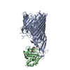
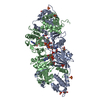
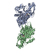
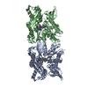
Crystal structure of phenamacril-bound F. graminearum myosin I
Components
Calmodulin
myosin I
Keywords
HYDROLASE / Myosin I / phenamcril / ATPase activity / fungicide / F. graminearum
Function / homology
Function and homology information
spindle pole body organization / central plaque of spindle pole body / actin cortical patch / myosin complex / cytoskeletal motor activity / enzyme regulator activity / actin binding / hydrolase activity / calcium ion binding / ATP binding / metal ion binding Similarity search - Function
Fungal myosin-I, SH3 domain / Single alpha-helices involved in coiled-coils or other helix-helix interfaces - #4820 / Class I myosin tail homology domain / Class I myosin, motor domain / Unconventional myosin tail, actin- and lipid-binding / Class I myosin tail homology (TH1) domain profile. / Myosin VI head, motor domain, U50 subdomain / : / Methane Monooxygenase Hydroxylase; Chain G, domain 1 - #530 / Ca2+ insensitive EF hand ...Fungal myosin-I, SH3 domain / Single alpha-helices involved in coiled-coils or other helix-helix interfaces - #4820 / Class I myosin tail homology domain / Class I myosin, motor domain / Unconventional myosin tail, actin- and lipid-binding / Class I myosin tail homology (TH1) domain profile. / Myosin VI head, motor domain, U50 subdomain / : / Methane Monooxygenase Hydroxylase; Chain G, domain 1 - #530 / Ca2+ insensitive EF hand / Myosin motor domain profile. / Myosin head, motor domain / Myosin head (motor domain) / Myosin. Large ATPases. / Kinesin motor domain superfamily / EF-hand / Recoverin; domain 1 / Single alpha-helices involved in coiled-coils or other helix-helix interfaces / SH3 domain / Methane Monooxygenase Hydroxylase; Chain G, domain 1 / Four Helix Bundle (Hemerythrin (Met), subunit A) / EF-hand domain pair / EF-hand, calcium binding motif / Src homology 3 domains / SH3-like domain superfamily / Src homology 3 (SH3) domain profile. / SH3 domain / EF-Hand 1, calcium-binding site / EF-hand calcium-binding domain. / EF-hand calcium-binding domain profile. / EF-hand domain / EF-hand domain pair / Up-down Bundle / P-loop containing nucleoside triphosphate hydrolase / Orthogonal Bundle / Mainly Alpha Similarity search - Domain/homology
 Movie
Movie Controller


 Open data
 Basic information
 Components
 Keywords
 Function and homology information
 Gibberella zeae (fungus)
 X-RAY DIFFRACTION /
  Authors
 China, 2items
  Citation
 Structure visualization
 Downloads & links
 Other downloads


 PDBj
PDBj


 Assembly
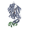

 Spodoptera frugiperda (fall armyworm) / References: UniProt: A0A098D3M4, UniProt: I1RCT2*PLUS


 Mass: 24.305 Da / Num. of mol.: 1 / Source method: obtained synthetically / Formula: Mg
 Mass: 216.236 Da / Num. of mol.: 1 / Source method: obtained synthetically / Formula: C12H12N2O2 / Feature type: SUBJECT OF INVESTIGATION
 Mass: 523.247 Da / Num. of mol.: 1 / Source method: obtained synthetically / Formula: C10H16N5O12P3S / Comment: ATP-gamma-S, energy-carrying molecule analogue*YM
 Sample preparation
 / Beamline: 21-ID-D / Wavelength: 1.12 Å
 Processing