 Movie
Movie Controller


+ Open data

- Basic information
| Entry | Database: PDB / ID: 4z8u | |||||||||||||||
|---|---|---|---|---|---|---|---|---|---|---|---|---|---|---|---|---|
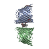
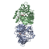
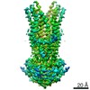
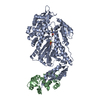
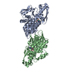
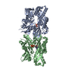
| Title | CRYSTAL STRUCTURE OF AvrRxo1-ORF1:-ORF2 WITH ATP | |||||||||||||||
 Components Components |
| |||||||||||||||
 Keywords Keywords | PROTEIN BINDING / AVRRXO1-ORF2 AVRRXO1-ORF1 AVRRXO1 AVRRXO1 REQUIRED CHAPERONE 1 / EFFECTOR PROTEINS AND MOLECULAR CHAPERONE / ATP | |||||||||||||||
| Function / homology | P-loop containing nucleoside triphosphate hydrolase / ACETATE ION / AvrRxo1-ORF1 / AvrRxo1-ORF2 Function and homology information Function and homology information | |||||||||||||||
| Biological species |  Xanthomonas oryzae pv. oryzicola (bacteria) Xanthomonas oryzae pv. oryzicola (bacteria) | |||||||||||||||
| Method |  X-RAY DIFFRACTION / SYNCHROTRON / MOLECULAR REPLACEMENT / Resolution: 1.65 Å X-RAY DIFFRACTION / SYNCHROTRON / MOLECULAR REPLACEMENT / Resolution: 1.65 Å | |||||||||||||||
 Authors Authors | Han, Q. / Zhou, C. / Wu, S. / Liu, Y. / Yang, Z. / Miao, J. / Triplett, L. / Cheng, Q. / Tokuhisa, J. / Deblais, L. ...Han, Q. / Zhou, C. / Wu, S. / Liu, Y. / Yang, Z. / Miao, J. / Triplett, L. / Cheng, Q. / Tokuhisa, J. / Deblais, L. / Robinson, H. / Leach, J.E. / Li, J. / Zhao, B. | |||||||||||||||
| Funding support |  United States, United States,  China, 4items China, 4items
| |||||||||||||||
 Citation Citation | Journal: Structure / Year: 2015 Title: Crystal Structure of Xanthomonas AvrRxo1-ORF1, a Type III Effector with a Polynucleotide Kinase Domain, and Its Interactor AvrRxo1-ORF2. Authors: Han, Q. / Zhou, C. / Wu, S. / Liu, Y. / Triplett, L. / Miao, J. / Tokuhisa, J. / Deblais, L. / Robinson, H. / Leach, J.E. / Li, J. / Zhao, B. | |||||||||||||||
| History |
|
- Structure visualization
| Structure viewer | Molecule: MolmilJmol/JSmol |
|---|
- Downloads & links
-Download
| PDBx/mmCIF format | 4z8u.cif.gz | 107.9 KB | Display | PDBx/mmCIF format |
|---|---|---|---|---|
| PDB format | pdb4z8u.ent.gz | 79.9 KB | Display | PDB format |
| PDBx/mmJSON format | 4z8u.json.gz | Tree view | PDBx/mmJSON format | |
| Others |  Other downloads Other downloads |
-Validation report
| Arichive directory | https://data.pdbj.org/pub/pdb/validation_reports/z8/4z8uftp://data.pdbj.org/pub/pdb/validation_reports/z8/4z8u | HTTPS FTP |
|---|
-Related structure data
| Related structure data |  4z8qC  4z8tC  4z8vC 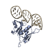 4pdbS C: citing same article ( S: Starting model for refinement |
|---|---|
| Similar structure data |


-Links
 PDBj
PDBj- Assembly
| Deposited unit | 
| ||||||||
|---|---|---|---|---|---|---|---|---|---|
| 1 | 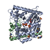
| ||||||||
| Unit cell |
| ||||||||
| Components on special symmetry positions |
|
-Components
-Protein , 2 types, 2 molecules AB
| #1: Protein | Mass: 36303.531 Da / Num. of mol.: 1 / Fragment: UNP RESIDUES 90-421 Source method: isolated from a genetically manipulated source Source: (gene. exp.) Xanthomonas oryzae pv. oryzicola (bacteria)Production host: |
|---|---|
| #2: Protein | Mass: 11523.586 Da / Num. of mol.: 1 / Fragment: UNP RESIDUES 1-98 Source method: isolated from a genetically manipulated source Source: (gene. exp.) Xanthomonas oryzae pv. oryzicola (bacteria)Production host: |
-Non-polymers , 4 types, 327 molecules 


| #3: Chemical | ChemComp-SO4 /  Mass: 96.063 Da / Num. of mol.: 5 / Source method: obtained synthetically / Formula: SO4 Mass: 96.063 Da / Num. of mol.: 5 / Source method: obtained synthetically / Formula: SO4#4: Chemical | ChemComp-GOL / |  Mass: 92.094 Da / Num. of mol.: 1 / Source method: obtained synthetically / Formula: C3H8O3 Mass: 92.094 Da / Num. of mol.: 1 / Source method: obtained synthetically / Formula: C3H8O3#5: Chemical | ChemComp-ACT / |  Mass: 59.044 Da / Num. of mol.: 1 / Source method: obtained synthetically / Formula: C2H3O2 Mass: 59.044 Da / Num. of mol.: 1 / Source method: obtained synthetically / Formula: C2H3O2#6: Water | ChemComp-HOH / | Mass: 18.015 Da / Num. of mol.: 320 / Source method: isolated from a natural source / Formula: H2O |
|---|
-Details
| Has protein modification | Y |
|---|
-Experimental details
-Experiment
| Experiment | Method: X-RAY DIFFRACTION / Number of used crystals: 1 |
|---|
- Sample preparation
| Crystal | Density Matthews: 2.65 Å3/Da / Density % sol: 53.65 % |
|---|---|
| Crystal grow | Temperature: 293 K / Method: vapor diffusion, hanging drop / pH: 5 Details: 1.6M (NH4)2SO4, 18% GLYCEROL, 8 MM DTT, 100 MM NA-CITRATE, 30 MM ATP AND 60 MM MAGNESIUM ACETATE, PH 5.0, VAPOR DIFFUSION, HANGING DROP, TEMPERATURE 293K PH range: 5 |
-Data collection
| Diffraction | Mean temperature: 100 K |
|---|---|
| Diffraction source | Source: SYNCHROTRON / Site: NSLS / Beamline: X29A / Wavelength: 1.075 Å |
| Detector | Type: ADSC QUANTUM 315r / Detector: CCD / Date: Mar 21, 2012 |
| Radiation | Monochromator: SI 111 CHANNEL / Protocol: SINGLE WAVELENGTH / Monochromatic (M) / Laue (L): M / Scattering type: x-ray |
| Radiation wavelength | Wavelength: 1.075 Å / Relative weight: 1 |
| Reflection | Resolution: 1.64→50 Å / Num. obs: 58250 / % possible obs: 98.6 % / Redundancy: 7.9 % / Rmerge(I) obs: 0.087 / Rsym value: 0.09 / Net I/σ(I): 20.1 |
| Reflection shell | Resolution: 1.64→1.7 Å / Redundancy: 5.5 % / Rmerge(I) obs: 0.618 / Mean I/σ(I) obs: 1.5 / Rsym value: 0.771 / % possible all: 85.7 |
- Processing
| Software |
| ||||||||||||||||||||||||||||||||||||||||||||||||||||||||||||||||||||||||||||||||||||||||||||||||||||||||||||||||||||||||||||||||||||||||||||||||||||||||||||||||||||||||||||||||||||||
|---|---|---|---|---|---|---|---|---|---|---|---|---|---|---|---|---|---|---|---|---|---|---|---|---|---|---|---|---|---|---|---|---|---|---|---|---|---|---|---|---|---|---|---|---|---|---|---|---|---|---|---|---|---|---|---|---|---|---|---|---|---|---|---|---|---|---|---|---|---|---|---|---|---|---|---|---|---|---|---|---|---|---|---|---|---|---|---|---|---|---|---|---|---|---|---|---|---|---|---|---|---|---|---|---|---|---|---|---|---|---|---|---|---|---|---|---|---|---|---|---|---|---|---|---|---|---|---|---|---|---|---|---|---|---|---|---|---|---|---|---|---|---|---|---|---|---|---|---|---|---|---|---|---|---|---|---|---|---|---|---|---|---|---|---|---|---|---|---|---|---|---|---|---|---|---|---|---|---|---|---|---|---|---|
| Refinement | Method to determine structure: MOLECULAR REPLACEMENT Starting model: 4PDB ENTRY Q5L Resolution: 1.65→45.82 Å / Cor.coef. Fo:Fc: 0.963 / Cor.coef. Fo:Fc free: 0.946 / Cross valid method: THROUGHOUT / ESU R: 0.098 / ESU R Free: 0.1 / Stereochemistry target values: MAXIMUM LIKELIHOOD
| ||||||||||||||||||||||||||||||||||||||||||||||||||||||||||||||||||||||||||||||||||||||||||||||||||||||||||||||||||||||||||||||||||||||||||||||||||||||||||||||||||||||||||||||||||||||
| Solvent computation | Ion probe radii: 0.8 Å / Shrinkage radii: 0.8 Å / VDW probe radii: 1.2 Å / Solvent model: MASK | ||||||||||||||||||||||||||||||||||||||||||||||||||||||||||||||||||||||||||||||||||||||||||||||||||||||||||||||||||||||||||||||||||||||||||||||||||||||||||||||||||||||||||||||||||||||
| Displacement parameters | Biso mean: 32.44 Å2
| ||||||||||||||||||||||||||||||||||||||||||||||||||||||||||||||||||||||||||||||||||||||||||||||||||||||||||||||||||||||||||||||||||||||||||||||||||||||||||||||||||||||||||||||||||||||
| Refinement step | Cycle: LAST / Resolution: 1.65→45.82 Å
| ||||||||||||||||||||||||||||||||||||||||||||||||||||||||||||||||||||||||||||||||||||||||||||||||||||||||||||||||||||||||||||||||||||||||||||||||||||||||||||||||||||||||||||||||||||||
| Refine LS restraints |
|