 Movie
Movie Controller


+ Open data

- Basic information
| Entry | Database: PDB / ID: 6ug2 | |||||||||
|---|---|---|---|---|---|---|---|---|---|---|
| Title | C2 symmetric peptide design number 1, Zappy, crystal form 2 | |||||||||
 Components Components | C2-1, Zappy, crystal form 2 | |||||||||
 Keywords Keywords | DE NOVO PROTEIN / cyclic peptide / 2-fold symmetric / L and D-amino acids | |||||||||
| Biological species | synthetic construct (others) | |||||||||
| Method |  X-RAY DIFFRACTION / SYNCHROTRON / AB INITIO PHASING / Resolution: 1.1 Å X-RAY DIFFRACTION / SYNCHROTRON / AB INITIO PHASING / Resolution: 1.1 Å | |||||||||
| Model details | S2 symmetric cyclic peptide | |||||||||
 Authors Authors | Mulligan, V.K. / Kang, C.S. / Antselovich, I. / Sawaya, M.R. / Yeates, T.O. / Baker, D. | |||||||||
| Funding support |  United States, 2items United States, 2items
| |||||||||
 Citation Citation | Journal: Protein Sci. / Year: 2020 Title: Computational design of mixed chirality peptide macrocycles with internal symmetry. Authors: Mulligan, V.K. / Kang, C.S. / Sawaya, M.R. / Rettie, S. / Li, X. / Antselovich, I. / Craven, T.W. / Watkins, A.M. / Labonte, J.W. / DiMaio, F. / Yeates, T.O. / Baker, D. | |||||||||
| History |
|
- Structure visualization
| Structure viewer | Molecule:  MolmilJmol/JSmol MolmilJmol/JSmol |
|---|
- Downloads & links
-Download
| PDBx/mmCIF format | 6ug2.cif.gz | 17.5 KB | Display | PDBx/mmCIF format |
|---|---|---|---|---|
| PDB format | pdb6ug2.ent.gz | 11.9 KB | Display | PDB format |
| PDBx/mmJSON format | 6ug2.json.gz | Tree view | PDBx/mmJSON format | |
| Others |  Other downloads Other downloads |
-Validation report
| Arichive directory | https://data.pdbj.org/pub/pdb/validation_reports/ug/6ug2ftp://data.pdbj.org/pub/pdb/validation_reports/ug/6ug2 | HTTPS FTP |
|---|
-Related structure data
| Related structure data | 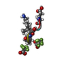 6ucxC 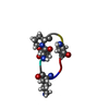 6ud9C 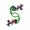 6udrC  6udwC  6udzC  6uf4C  6uf7C  6uf8C 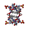 6uf9C 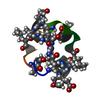 6ufaC  6ufuC 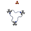 6ug3C 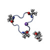 6ug6C  6ugbC 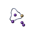 6ugcC C: citing same article ( |
|---|
-Links
 PDBj
PDBj
- Assembly
| Deposited unit | 
| ||||||||||
|---|---|---|---|---|---|---|---|---|---|---|---|
| 1 |
| ||||||||||
| Unit cell |
|
-Components
| #1: Protein/peptide | Mass: 1021.079 Da / Num. of mol.: 1 / Source method: obtained synthetically Details: Residue 1 is partially racemized. Both DSN and SER are present at this position. The racemization was an undesired side product of the peptide synthesis. Source: (synth.) synthetic construct (others) |
|---|---|
| #2: Chemical | ChemComp-CA /   Mass: 40.078 Da / Num. of mol.: 1 / Source method: obtained synthetically / Formula: Ca Mass: 40.078 Da / Num. of mol.: 1 / Source method: obtained synthetically / Formula: Ca |
| #3: Water | ChemComp-HOH /  Mass: 18.015 Da / Num. of mol.: 15 / Source method: isolated from a natural source / Formula: H2O Mass: 18.015 Da / Num. of mol.: 15 / Source method: isolated from a natural source / Formula: H2O |
| Compound details | Residue 1 is partially racemized. Both DSN and SER are present at this position. The racemization ...Residue 1 is partially racemized. Both DSN and SER are present at this position. The racemization was an undesired side product of the peptide synthesis. |
| Has ligand of interest | N |
| Has protein modification | Y |
-Experimental details
-Experiment
| Experiment | Method: X-RAY DIFFRACTION / Number of used crystals: 1 |
|---|
- Sample preparation
| Crystal | Density Matthews: 1.55 Å3/Da / Density % sol: 20.86 % Description: needle, 200 microns long and less than 5 microns thick |
|---|---|
| Crystal grow | Temperature: 298 K / Method: vapor diffusion, hanging drop / pH: 7.5 Details: 0.2 M calcium chloride, 0.1 M HEPES, pH 7.5, 28% (w/v) PEG 400 |
-Data collection
| Diffraction | Mean temperature: 100 K / Serial crystal experiment: N |
|---|---|
| Diffraction source | Source: SYNCHROTRON / Site: APS / Beamline: 24-ID-E / Wavelength: 0.97918 Å |
| Detector | Type: DECTRIS EIGER X 16M / Detector: PIXEL / Date: Feb 14, 2019 |
| Radiation | Monochromator: Si (111) / Protocol: SINGLE WAVELENGTH / Monochromatic (M) / Laue (L): M / Scattering type: x-ray |
| Radiation wavelength | Wavelength: 0.97918 Å / Relative weight: 1 |
| Reflection | Resolution: 1.1→19.734 Å / Num. obs: 4357 / % possible obs: 87.4 % / Redundancy: 4.753 % / Biso Wilson estimate: 11.43 Å2 / Rmerge(I) obs: 0.25 / Net I/σ(I): 4.08 |
| Reflection shell | Resolution: 1.1→1.13 Å / Redundancy: 4.06 % / Rmerge(I) obs: 0.898 / Mean I/σ(I) obs: 1.14 / Num. unique obs: 240 / % possible all: 66.9 |
- Processing
| Software |
| ||||||||||||||||||||||||||||
|---|---|---|---|---|---|---|---|---|---|---|---|---|---|---|---|---|---|---|---|---|---|---|---|---|---|---|---|---|---|
| Refinement | Method to determine structure: AB INITIO PHASING / Resolution: 1.1→19.73 Å / SU ML: 0.077 / Cross valid method: FREE R-VALUE / σ(F): 1.36 / Phase error: 17.01
| ||||||||||||||||||||||||||||
| Solvent computation | Shrinkage radii: 0.9 Å / VDW probe radii: 1.11 Å | ||||||||||||||||||||||||||||
| Displacement parameters | Biso mean: 11.18 Å2 | ||||||||||||||||||||||||||||
| Refinement step | Cycle: LAST / Resolution: 1.1→19.73 Å
| ||||||||||||||||||||||||||||
| Refine LS restraints |
| ||||||||||||||||||||||||||||
| LS refinement shell |
|
