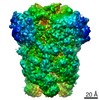
 Movie
Movie Controller


+ Open data

- Basic information
| Entry | Database: PDB / ID: 6u7e | |||||||||
|---|---|---|---|---|---|---|---|---|---|---|
| Title | HCoV-229E RBD Class III in complex with human APN | |||||||||
 Components Components |
| |||||||||
 Keywords Keywords | HYDROLASE/VIRAL PROTEIN / CoV / coronavirus / 229E / spike glycoprotein / APN / S-protein / HYDROLASE-VIRAL PROTEIN complex | |||||||||
| Function / homology |  Function and homology information Function and homology informationmembrane alanyl aminopeptidase / alanyl aminopeptidase activity / peptide catabolic process / metalloaminopeptidase activity / endoplasmic reticulum-Golgi intermediate compartment / aminopeptidase activity / Metabolism of Angiotensinogen to Angiotensins / angiotensin maturation / secretory granule membrane / metallopeptidase activity ...membrane alanyl aminopeptidase / alanyl aminopeptidase activity / peptide catabolic process / metalloaminopeptidase activity / endoplasmic reticulum-Golgi intermediate compartment / aminopeptidase activity / Metabolism of Angiotensinogen to Angiotensins / angiotensin maturation / secretory granule membrane / metallopeptidase activity / virus receptor activity / signaling receptor activity / angiogenesis / host cell endoplasmic reticulum-Golgi intermediate compartment membrane / cell differentiation / receptor-mediated virion attachment to host cell / endocytosis involved in viral entry into host cell / fusion of virus membrane with host plasma membrane / external side of plasma membrane / lysosomal membrane / fusion of virus membrane with host endosome membrane / viral envelope / Neutrophil degranulation / virion membrane / : / extracellular exosome / zinc ion binding / membrane / plasma membrane Similarity search - Function | |||||||||
| Biological species |  Homo sapiens (human) Homo sapiens (human) Human coronavirus 229E Human coronavirus 229E | |||||||||
| Method |  X-RAY DIFFRACTION / SYNCHROTRON / MOLECULAR REPLACEMENT / Resolution: 3 Å X-RAY DIFFRACTION / SYNCHROTRON / MOLECULAR REPLACEMENT / Resolution: 3 Å | |||||||||
 Authors Authors | Tomlinson, A.C.A. / Li, Z. / Rini, J.M. | |||||||||
| Funding support |  Canada, 1items Canada, 1items
| |||||||||
 Citation Citation | Journal: Elife / Year: 2019 Title: The human coronavirus HCoV-229E S-protein structure and receptor binding. Authors: Zhijie Li / Aidan Ca Tomlinson / Alan Hm Wong / Dongxia Zhou / Marc Desforges / Pierre J Talbot / Samir Benlekbir / John L Rubinstein / James M Rini / Abstract: The coronavirus S-protein mediates receptor binding and fusion of the viral and host cell membranes. In HCoV-229E, its receptor binding domain (RBD) shows extensive sequence variation but how S- ...The coronavirus S-protein mediates receptor binding and fusion of the viral and host cell membranes. In HCoV-229E, its receptor binding domain (RBD) shows extensive sequence variation but how S-protein function is maintained is not understood. Reported are the X-ray crystal structures of Class III-V RBDs in complex with human aminopeptidase N (hAPN), as well as the electron cryomicroscopy structure of the 229E S-protein. The structures show that common core interactions define the specificity for hAPN and that the peripheral RBD sequence variation is accommodated by loop plasticity. The results provide insight into immune evasion and the cross-species transmission of 229E and related coronaviruses. We also find that the 229E S-protein can expose a portion of its helical core to solvent. This is undoubtedly facilitated by hydrophilic subunit interfaces that we show are conserved among coronaviruses. These interfaces likely play a role in the S-protein conformational changes associated with membrane fusion. | |||||||||
| History |
|
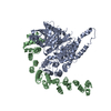
- Structure visualization
| Structure viewer | Molecule: MolmilJmol/JSmol |
|---|
- Downloads & links
-Download
| PDBx/mmCIF format | 6u7e.cif.gz | 834.9 KB | Display | PDBx/mmCIF format |
|---|---|---|---|---|
| PDB format | pdb6u7e.ent.gz | 692 KB | Display | PDB format |
| PDBx/mmJSON format | 6u7e.json.gz | Tree view | PDBx/mmJSON format | |
| Others |  Other downloads Other downloads |
-Validation report
| Arichive directory | https://data.pdbj.org/pub/pdb/validation_reports/u7/6u7eftp://data.pdbj.org/pub/pdb/validation_reports/u7/6u7e | HTTPS FTP |
|---|
-Related structure data
| Related structure data |  6u7fC  6u7gC  6u7hC  4fyqS C: citing same article ( S: Starting model for refinement |
|---|---|
| Similar structure data | |
| Experimental dataset #1 | Data reference: 10.18430/m36u7e / Data set type: diffraction image data |


-Links
 PDBj
PDBj


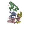
- Assembly
| Deposited unit | 
| ||||||||
|---|---|---|---|---|---|---|---|---|---|
| 1 | 
| ||||||||
| 2 | 
| ||||||||
| Unit cell |
|
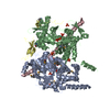
-Components
-Protein , 2 types, 4 molecules ABCD
| #1: Protein | Mass: 103522.031 Da / Num. of mol.: 2 / Fragment: ectodomain (UNP residues 66-967) Source method: isolated from a genetically manipulated source Source: (gene. exp.) Homo sapiens (human) / Gene: ANPEP, APN, CD13, PEPN / Plasmid: PB-T-PAF / Cell line (production host): HEK293S GnT1-minus / Production host: Homo sapiens (human) / References: UniProt: P15144, membrane alanyl aminopeptidase#2: Protein | Mass: 15664.968 Da / Num. of mol.: 2 Source method: isolated from a genetically manipulated source Source: (gene. exp.) Human coronavirus 229E / Plasmid: PB-T-PAF / Cell line (production host): HEK293S GnT1-minus / Production host: Homo sapiens (human) / References: UniProt: Q1HVL8*PLUS |
|---|
-Sugars , 3 types, 16 molecules 
| #3: Polysaccharide | Source method: isolated from a genetically manipulated source #4: Polysaccharide | Source method: isolated from a genetically manipulated source #6: Sugar | ChemComp-NAG /  Type: D-saccharide, beta linking / Mass: 221.208 Da / Num. of mol.: 12 Type: D-saccharide, beta linking / Mass: 221.208 Da / Num. of mol.: 12Source method: isolated from a genetically manipulated source Formula: C8H15NO6 |
|---|
-Non-polymers , 2 types, 7 molecules 

| #5: Chemical |  Mass: 65.409 Da / Num. of mol.: 2 / Source method: obtained synthetically / Formula: Zn Mass: 65.409 Da / Num. of mol.: 2 / Source method: obtained synthetically / Formula: Zn#7: Water | ChemComp-HOH / | Mass: 18.015 Da / Num. of mol.: 5 / Source method: isolated from a natural source / Formula: H2O |
|---|
-Details
| Has ligand of interest | N |
|---|---|
| Has protein modification | Y |
-Experimental details
-Experiment
| Experiment | Method: X-RAY DIFFRACTION / Number of used crystals: 1 |
|---|
- Sample preparation
| Crystal | Density Matthews: 3.07 Å3/Da / Density % sol: 59.97 % |
|---|---|
| Crystal grow | Temperature: 298 K / Method: vapor diffusion, hanging drop / pH: 6.5 Details: 9% PEG8000, 1 mM oxidized glutathione, 1 mM reduced glutathione, 5% glycerol, 100 mM MES, pH 6.5 |
-Data collection
| Diffraction | Mean temperature: 100 K / Serial crystal experiment: N | ||||||||||||||||||||||||||||||||||||||||||||||||||||||||||||||||||||||||||||||||||||||||||||||||||||||||||||||||||||||||||||||||||||||||||||||||||||||||||||||||||||||||||||||||||||||||||||||||||||||||||||||||||
|---|---|---|---|---|---|---|---|---|---|---|---|---|---|---|---|---|---|---|---|---|---|---|---|---|---|---|---|---|---|---|---|---|---|---|---|---|---|---|---|---|---|---|---|---|---|---|---|---|---|---|---|---|---|---|---|---|---|---|---|---|---|---|---|---|---|---|---|---|---|---|---|---|---|---|---|---|---|---|---|---|---|---|---|---|---|---|---|---|---|---|---|---|---|---|---|---|---|---|---|---|---|---|---|---|---|---|---|---|---|---|---|---|---|---|---|---|---|---|---|---|---|---|---|---|---|---|---|---|---|---|---|---|---|---|---|---|---|---|---|---|---|---|---|---|---|---|---|---|---|---|---|---|---|---|---|---|---|---|---|---|---|---|---|---|---|---|---|---|---|---|---|---|---|---|---|---|---|---|---|---|---|---|---|---|---|---|---|---|---|---|---|---|---|---|---|---|---|---|---|---|---|---|---|---|---|---|---|---|---|---|---|
| Diffraction source | Source: SYNCHROTRON / Site: CLSI / Beamline: 08ID-1 / Wavelength: 0.97949 Å | ||||||||||||||||||||||||||||||||||||||||||||||||||||||||||||||||||||||||||||||||||||||||||||||||||||||||||||||||||||||||||||||||||||||||||||||||||||||||||||||||||||||||||||||||||||||||||||||||||||||||||||||||||
| Detector | Type: RAYONIX MX-300 / Detector: CCD / Date: Jan 21, 2017 | ||||||||||||||||||||||||||||||||||||||||||||||||||||||||||||||||||||||||||||||||||||||||||||||||||||||||||||||||||||||||||||||||||||||||||||||||||||||||||||||||||||||||||||||||||||||||||||||||||||||||||||||||||
| Radiation | Monochromator: double crystal Si(111) / Protocol: SINGLE WAVELENGTH / Monochromatic (M) / Laue (L): M / Scattering type: x-ray | ||||||||||||||||||||||||||||||||||||||||||||||||||||||||||||||||||||||||||||||||||||||||||||||||||||||||||||||||||||||||||||||||||||||||||||||||||||||||||||||||||||||||||||||||||||||||||||||||||||||||||||||||||
| Radiation wavelength | Wavelength: 0.97949 Å / Relative weight: 1 | ||||||||||||||||||||||||||||||||||||||||||||||||||||||||||||||||||||||||||||||||||||||||||||||||||||||||||||||||||||||||||||||||||||||||||||||||||||||||||||||||||||||||||||||||||||||||||||||||||||||||||||||||||
| Reflection | Resolution: 2.99→48.092 Å / Num. obs: 58215 / % possible obs: 99.8 % / Redundancy: 3.642 % / Biso Wilson estimate: 81.905 Å2 / CC1/2: 0.998 / Rmerge(I) obs: 0.071 / Rrim(I) all: 0.083 / Χ2: 1.024 / Net I/σ(I): 11.06 / Num. measured all: 212045 / Scaling rejects: 12 | ||||||||||||||||||||||||||||||||||||||||||||||||||||||||||||||||||||||||||||||||||||||||||||||||||||||||||||||||||||||||||||||||||||||||||||||||||||||||||||||||||||||||||||||||||||||||||||||||||||||||||||||||||
| Reflection shell | Diffraction-ID: 1
|
- Processing
| Software |
| ||||||||||||||||||||||||||||||||||||||||||||||||||||||||||||||||||||||||||||||||||||||||||||||||||||||||||||||||||||||||||||||||||||
|---|---|---|---|---|---|---|---|---|---|---|---|---|---|---|---|---|---|---|---|---|---|---|---|---|---|---|---|---|---|---|---|---|---|---|---|---|---|---|---|---|---|---|---|---|---|---|---|---|---|---|---|---|---|---|---|---|---|---|---|---|---|---|---|---|---|---|---|---|---|---|---|---|---|---|---|---|---|---|---|---|---|---|---|---|---|---|---|---|---|---|---|---|---|---|---|---|---|---|---|---|---|---|---|---|---|---|---|---|---|---|---|---|---|---|---|---|---|---|---|---|---|---|---|---|---|---|---|---|---|---|---|---|---|
| Refinement | Method to determine structure: MOLECULAR REPLACEMENT Starting model: PDB entry 4FYQ Resolution: 3→48.092 Å / SU ML: 0.4 / Cross valid method: THROUGHOUT / σ(F): 1.36 / Phase error: 28.05 / Stereochemistry target values: ML
| ||||||||||||||||||||||||||||||||||||||||||||||||||||||||||||||||||||||||||||||||||||||||||||||||||||||||||||||||||||||||||||||||||||
| Solvent computation | Shrinkage radii: 0.9 Å / VDW probe radii: 1.11 Å / Solvent model: FLAT BULK SOLVENT MODEL | ||||||||||||||||||||||||||||||||||||||||||||||||||||||||||||||||||||||||||||||||||||||||||||||||||||||||||||||||||||||||||||||||||||
| Displacement parameters | Biso max: 227.29 Å2 / Biso mean: 97.7358 Å2 / Biso min: 46.4 Å2 | ||||||||||||||||||||||||||||||||||||||||||||||||||||||||||||||||||||||||||||||||||||||||||||||||||||||||||||||||||||||||||||||||||||
| Refinement step | Cycle: final / Resolution: 3→48.092 Å
| ||||||||||||||||||||||||||||||||||||||||||||||||||||||||||||||||||||||||||||||||||||||||||||||||||||||||||||||||||||||||||||||||||||
| LS refinement shell | Refine-ID: X-RAY DIFFRACTION / Rfactor Rfree error: 0
| ||||||||||||||||||||||||||||||||||||||||||||||||||||||||||||||||||||||||||||||||||||||||||||||||||||||||||||||||||||||||||||||||||||
| Refinement TLS params. | Method: refined / Origin x: -24.614 Å / Origin y: -18.6671 Å / Origin z: -38.2449 Å
| ||||||||||||||||||||||||||||||||||||||||||||||||||||||||||||||||||||||||||||||||||||||||||||||||||||||||||||||||||||||||||||||||||||
| Refinement TLS group |
|
