 Movie
Movie Controller

[English] 日本語
 Yorodumi
- PDB-5bv1: Crystal Structure of a Vps33-Vps16 Complex from Chaetomium thermo... -
+ Open data

- Basic information
| Entry | Database: PDB / ID: 5bv1 | ||||||
|---|---|---|---|---|---|---|---|
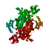
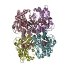
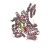
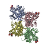
| Title | Crystal Structure of a Vps33-Vps16 Complex from Chaetomium thermophilum | ||||||
 Components Components |
| ||||||
 Keywords Keywords | TRANSPORT PROTEIN / Membrane trafficking / SM protein / HOPS complex / thermophile | ||||||
| Function / homology |  Function and homology information Function and homology informationHOPS complex / vacuole fusion, non-autophagic / endosomal transport / ligase activity / vesicle-mediated transport / intracellular protein transport / actin binding / endosome Similarity search - Function | ||||||
| Biological species |  Chaetomium thermophilum (fungus) Chaetomium thermophilum (fungus) | ||||||
| Method |  X-RAY DIFFRACTION / SYNCHROTRON / MOLECULAR REPLACEMENT / molecular replacement / Resolution: 2.902 Å X-RAY DIFFRACTION / SYNCHROTRON / MOLECULAR REPLACEMENT / molecular replacement / Resolution: 2.902 Å | ||||||
 Authors Authors | Baker, R.W. / Jeffrey, P.D. / Hughson, F.M. | ||||||
| Funding support |  United States, 1items United States, 1items
| ||||||
 Citation Citation | Journal: Science / Year: 2015 Title: A direct role for the Sec1/Munc18-family protein Vps33 as a template for SNARE assembly. Authors: Baker, R.W. / Jeffrey, P.D. / Zick, M. / Phillips, B.P. / Wickner, W.T. / Hughson, F.M. #1: Journal: Plos One / Year: 2013Title: Crystal Structures of the Sec1/Munc18 (SM) Protein Vps33, Alone and Bound to the Homotypic Fusion and Vacuolar Protein Sorting (HOPS) Subunit Vps16* Authors: Baker, R.W. / Jeffrey, P.D. / Hughson, F.M. | ||||||
| History |
|
- Structure visualization
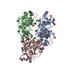
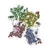
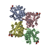
| Structure viewer | Molecule: MolmilJmol/JSmol |
|---|
- Downloads & links
-Download
| PDBx/mmCIF format | 5bv1.cif.gz | 363 KB | Display | PDBx/mmCIF format |
|---|---|---|---|---|
| PDB format | pdb5bv1.ent.gz | 290.8 KB | Display | PDB format |
| PDBx/mmJSON format | 5bv1.json.gz | Tree view | PDBx/mmJSON format | |
| Others |  Other downloads Other downloads |
-Validation report
| Arichive directory | https://data.pdbj.org/pub/pdb/validation_reports/bv/5bv1ftp://data.pdbj.org/pub/pdb/validation_reports/bv/5bv1 | HTTPS FTP |
|---|
-Related structure data
| Related structure data |  5buzC  5bv0C  4kmoS S: Starting model for refinement C: citing same article ( |
|---|---|
| Similar structure data |

-Links
 PDBj
PDBj


- Assembly
| Deposited unit | 
| |||||||||||||||||||||||||||||||||||||||||||||||||||||||||||||||
|---|---|---|---|---|---|---|---|---|---|---|---|---|---|---|---|---|---|---|---|---|---|---|---|---|---|---|---|---|---|---|---|---|---|---|---|---|---|---|---|---|---|---|---|---|---|---|---|---|---|---|---|---|---|---|---|---|---|---|---|---|---|---|---|---|
| 1 | 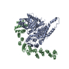
| |||||||||||||||||||||||||||||||||||||||||||||||||||||||||||||||
| 2 | 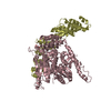
| |||||||||||||||||||||||||||||||||||||||||||||||||||||||||||||||
| Unit cell |
| |||||||||||||||||||||||||||||||||||||||||||||||||||||||||||||||
| Noncrystallographic symmetry (NCS) | NCS domain:
NCS domain segments: Component-ID: 1 / Beg auth comp-ID: ALA / Beg label comp-ID: ALA
NCS ensembles :
|
-Components
| #1: Protein | Mass: 73990.117 Da / Num. of mol.: 2 / Fragment: unp residues 139-806 Source method: isolated from a genetically manipulated source Source: (gene. exp.) Chaetomium thermophilum (strain DSM 1495 / CBS 144.50 / IMI 039719) (fungus)Strain: DSM 1495 / CBS 144.50 / IMI 039719 / Gene: CTHT_0057760 / Plasmid: pQLinkH / Production host:  #2: Protein | Mass: 37912.211 Da / Num. of mol.: 2 / Fragment: unp residues 503-816 / Mutation: L672V Source method: isolated from a genetically manipulated source Source: (gene. exp.) Chaetomium thermophilum (fungus) / Strain: DSM 1495 / CBS 144.50 / IMI 039719 / Gene: CTHT_0026760 / Plasmid: pQLinkH / Production host: #3: Chemical |   Mass: 134.087 Da / Num. of mol.: 2 / Source method: obtained synthetically / Formula: C4H6O5 Mass: 134.087 Da / Num. of mol.: 2 / Source method: obtained synthetically / Formula: C4H6O5Sequence details | Authors have indicated that the sequence for the UNProt entries G0S6M7_CHATD and G0SCM5_CHATD are not correct | |
|---|
-Experimental details
-Experiment
| Experiment | Method: X-RAY DIFFRACTION / Number of used crystals: 1 |
|---|
- Sample preparation
| Crystal | Density Matthews: 2.76 Å3/Da / Density % sol: 60.86 % |
|---|---|
| Crystal grow | Temperature: 293 K / Method: vapor diffusion, sitting drop / pH: 7 / Details: HEPES buffer, 8-10% w/v PEG 5000 monomethyl ether |
-Data collection
| Diffraction | Mean temperature: 100 K | |||||||||||||||||||||
|---|---|---|---|---|---|---|---|---|---|---|---|---|---|---|---|---|---|---|---|---|---|---|
| Diffraction source | Source: SYNCHROTRON / Site: NSLS / Beamline: X29A / Wavelength: 1.075 Å | |||||||||||||||||||||
| Detector | Type: ADSC QUANTUM 315r / Detector: CCD / Date: Apr 28, 2014 | |||||||||||||||||||||
| Radiation | Monochromator: Silicon / Protocol: SINGLE WAVELENGTH / Monochromatic (M) / Laue (L): M / Scattering type: x-ray | |||||||||||||||||||||
| Radiation wavelength | Wavelength: 1.075 Å / Relative weight: 1 | |||||||||||||||||||||
| Reflection | Resolution: 2.902→159.902 Å / Num. obs: 53085 / % possible obs: 99 % / Observed criterion σ(I): -3 / Redundancy: 5.7 % / Biso Wilson estimate: 65.34 Å2 / Rmerge(I) obs: 0.085 / Net I/σ(I): 16.9 / Num. measured all: 300496 | |||||||||||||||||||||
| Reflection shell | Diffraction-ID: 1 / Rejects: _
|
-Phasing
| Phasing | Method: molecular replacement |
|---|
- Processing
| Software |
| ||||||||||||||||||||||||||||||||||||||||||||||||||||||||||||||||||||||||||||||||||||||||||||||||||||||||||||||||||||||||||||||||||||||||||||
|---|---|---|---|---|---|---|---|---|---|---|---|---|---|---|---|---|---|---|---|---|---|---|---|---|---|---|---|---|---|---|---|---|---|---|---|---|---|---|---|---|---|---|---|---|---|---|---|---|---|---|---|---|---|---|---|---|---|---|---|---|---|---|---|---|---|---|---|---|---|---|---|---|---|---|---|---|---|---|---|---|---|---|---|---|---|---|---|---|---|---|---|---|---|---|---|---|---|---|---|---|---|---|---|---|---|---|---|---|---|---|---|---|---|---|---|---|---|---|---|---|---|---|---|---|---|---|---|---|---|---|---|---|---|---|---|---|---|---|---|---|---|
| Refinement | Method to determine structure: MOLECULAR REPLACEMENT Starting model: 4KMO Resolution: 2.902→61.592 Å / SU ML: 0.47 / Cross valid method: THROUGHOUT / σ(F): 1.37 / Phase error: 30.6 / Stereochemistry target values: ML
| ||||||||||||||||||||||||||||||||||||||||||||||||||||||||||||||||||||||||||||||||||||||||||||||||||||||||||||||||||||||||||||||||||||||||||||
| Solvent computation | Shrinkage radii: 0.9 Å / VDW probe radii: 1.11 Å / Solvent model: FLAT BULK SOLVENT MODEL | ||||||||||||||||||||||||||||||||||||||||||||||||||||||||||||||||||||||||||||||||||||||||||||||||||||||||||||||||||||||||||||||||||||||||||||
| Displacement parameters | Biso max: 174.73 Å2 / Biso mean: 66.5912 Å2 / Biso min: 20.98 Å2 | ||||||||||||||||||||||||||||||||||||||||||||||||||||||||||||||||||||||||||||||||||||||||||||||||||||||||||||||||||||||||||||||||||||||||||||
| Refinement step | Cycle: final / Resolution: 2.902→61.592 Å
| ||||||||||||||||||||||||||||||||||||||||||||||||||||||||||||||||||||||||||||||||||||||||||||||||||||||||||||||||||||||||||||||||||||||||||||
| Refine LS restraints |
| ||||||||||||||||||||||||||||||||||||||||||||||||||||||||||||||||||||||||||||||||||||||||||||||||||||||||||||||||||||||||||||||||||||||||||||
| Refine LS restraints NCS |
| ||||||||||||||||||||||||||||||||||||||||||||||||||||||||||||||||||||||||||||||||||||||||||||||||||||||||||||||||||||||||||||||||||||||||||||
| LS refinement shell | Refine-ID: X-RAY DIFFRACTION / Total num. of bins used: 19
|
