Movie
Movie Controller

[English] 日本語
 Yorodumi
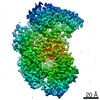
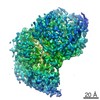
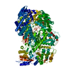
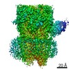
- PDB-6u1x: Structure of the Vesicular Stomatitis Virus L Protein in Complex ... -
+ Open data

- Basic information
| Entry | Database: PDB / ID: 6u1x | ||||||
|---|---|---|---|---|---|---|---|
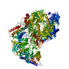
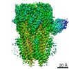
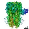
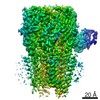
| Title | Structure of the Vesicular Stomatitis Virus L Protein in Complex with Its Phosphoprotein Cofactor (3.0 A resolution) | ||||||
 Components Components |
| ||||||
 Keywords Keywords | VIRAL PROTEIN / Vesicular stomatitis virus / RNA-dependent RNA polymerase / L protein / P protein / phosphoprotein / single particle analysis / transcription / replication / virus / viral / RdRp / PRNTase / nonsegmented negative-sense RNA viruses | ||||||
| Function / homology |  Function and homology information Function and homology informationGDP polyribonucleotidyltransferase / RNA folding chaperone / phosphorylation / negative stranded viral RNA replication / viral transcription / Hydrolases; Acting on acid anhydrides; In phosphorus-containing anhydrides / viral genome replication / virion component / host cell cytoplasm / mRNA 5'-cap (guanine-N7-)-methyltransferase activity ...GDP polyribonucleotidyltransferase / RNA folding chaperone / phosphorylation / negative stranded viral RNA replication / viral transcription / Hydrolases; Acting on acid anhydrides; In phosphorus-containing anhydrides / viral genome replication / virion component / host cell cytoplasm / mRNA 5'-cap (guanine-N7-)-methyltransferase activity / RNA-directed RNA polymerase / RNA-directed RNA polymerase activity / GTPase activity / ATP binding / metal ion binding Similarity search - Function | ||||||
| Biological species |  Vesicular stomatitis Indiana virus Vesicular stomatitis Indiana virus | ||||||
| Method | ELECTRON MICROSCOPY / single particle reconstruction / cryo EM / Resolution: 3 Å | ||||||
 Authors Authors | Jenni, S. / Bloyet, L.M. / Dias-Avalos, R. / Liang, B. / Wheelman, S.P.J. / Grigorieff, N. / Harrison, S.C. | ||||||
| Funding support |  United States, 1items United States, 1items
| ||||||
 Citation Citation | Journal: Cell Rep / Year: 2020 Title: Structure of the Vesicular Stomatitis Virus L Protein in Complex with Its Phosphoprotein Cofactor. Authors: Simon Jenni / Louis-Marie Bloyet / Ruben Diaz-Avalos / Bo Liang / Sean P J Whelan / Nikolaus Grigorieff / Stephen C Harrison / Abstract: The large (L) proteins of non-segmented, negative-strand RNA viruses are multifunctional enzymes that produce capped, methylated, and polyadenylated mRNA and replicate the viral genome. A ...The large (L) proteins of non-segmented, negative-strand RNA viruses are multifunctional enzymes that produce capped, methylated, and polyadenylated mRNA and replicate the viral genome. A phosphoprotein (P), required for efficient RNA-dependent RNA polymerization from the viral ribonucleoprotein (RNP) template, regulates the function and conformation of the L protein. We report the structure of vesicular stomatitis virus L in complex with its P cofactor determined by electron cryomicroscopy at 3.0 Å resolution, enabling us to visualize bound segments of P. The contacts of three P segments with multiple L domains show how P induces a closed, compact, initiation-competent conformation. Binding of P to L positions its N-terminal domain adjacent to a putative RNA exit channel for efficient encapsidation of newly synthesized genomes with the nucleoprotein and orients its C-terminal domain to interact with an RNP template. The model shows that a conserved tryptophan in the priming loop can support the initiating 5' nucleotide. | ||||||
| History |
|
- Structure visualization
| Movie |
Movie viewer |
|---|---|
| Structure viewer | Molecule: MolmilJmol/JSmol |
- Downloads & links
-Download
| PDBx/mmCIF format | 6u1x.cif.gz | 709.8 KB | Display | PDBx/mmCIF format |
|---|---|---|---|---|
| PDB format | pdb6u1x.ent.gz | 573.6 KB | Display | PDB format |
| PDBx/mmJSON format | 6u1x.json.gz | Tree view | PDBx/mmJSON format | |
| Others |  Other downloads Other downloads |
-Validation report
| Arichive directory | https://data.pdbj.org/pub/pdb/validation_reports/u1/6u1xftp://data.pdbj.org/pub/pdb/validation_reports/u1/6u1x | HTTPS FTP |
|---|
-Related structure data
| Related structure data |  20614MC M: map data used to model this data C: citing same article ( |
|---|---|
| Similar structure data |
-Links
 PDBj
PDBj
- Assembly
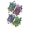
| Deposited unit | 
|
|---|---|
| 1 |
|
-Components
| #1: Protein | Mass: 241313.859 Da / Num. of mol.: 1 Source method: isolated from a genetically manipulated source Source: (gene. exp.) Vesicular stomatitis Indiana virus (strain San Juan)Strain: San Juan / Production host:   Spodoptera frugiperda (fall armyworm) Spodoptera frugiperda (fall armyworm)References: UniProt: P03523, RNA-directed RNA polymerase, mRNA (guanine-N7)-methyltransferase, GDP polyribonucleotidyltransferase, EC: 2.1.1.296 | ||
|---|---|---|---|
| #2: Protein | Mass: 29941.982 Da / Num. of mol.: 1 Source method: isolated from a genetically manipulated source Source: (gene. exp.) Vesicular stomatitis Indiana virus (strain San Juan)Strain: San Juan / Production host:  | ||
| #3: Chemical |   Mass: 65.409 Da / Num. of mol.: 2 / Source method: obtained synthetically / Formula: Zn / Feature type: SUBJECT OF INVESTIGATION Mass: 65.409 Da / Num. of mol.: 2 / Source method: obtained synthetically / Formula: Zn / Feature type: SUBJECT OF INVESTIGATIONHas ligand of interest | Y | |
-Experimental details
-Experiment
| Experiment | Method: ELECTRON MICROSCOPY |
|---|---|
| EM experiment | Aggregation state: PARTICLE / 3D reconstruction method: single particle reconstruction |
- Sample preparation
| Component | Name: VSV L:P / Type: COMPLEX Details: L protein (full-length) in complex with P protein (35-106) Entity ID: #1-#2 / Source: RECOMBINANT |
|---|---|
| Molecular weight | Value: 0.24 MDa / Experimental value: NO |
| Source (natural) | Organism: Vesicular stomatitis virus |
| Source (recombinant) | Organism: Spodoptera frugiperda (fall armyworm) |
| Buffer solution | pH: 7.4 |
| Specimen | Embedding applied: NO / Shadowing applied: NO / Staining applied: NO / Vitrification applied: YES |
| Vitrification | Cryogen name: ETHANE |
- Electron microscopy imaging
| Experimental equipment |  Model: Titan Krios / Image courtesy: FEI Company |
|---|---|
| Microscopy | Model: FEI TITAN KRIOS |
| Electron gun | Electron source:  FIELD EMISSION GUN / Accelerating voltage: 300 kV / Illumination mode: FLOOD BEAM FIELD EMISSION GUN / Accelerating voltage: 300 kV / Illumination mode: FLOOD BEAM |
| Electron lens | Mode: BRIGHT FIELD |
| Image recording | Average exposure time: 5 sec. / Electron dose: 100 e/Å2 / Detector mode: COUNTING / Film or detector model: GATAN K2 SUMMIT (4k x 4k) / Num. of grids imaged: 3 / Num. of real images: 3307 |
| Image scans | Sampling size: 5 µm / Width: 3838 / Height: 3710 / Movie frames/image: 50 / Used frames/image: 1-50 |
- Processing
| Software |
| ||||||||||||||||||||||||||||||||||||||||
|---|---|---|---|---|---|---|---|---|---|---|---|---|---|---|---|---|---|---|---|---|---|---|---|---|---|---|---|---|---|---|---|---|---|---|---|---|---|---|---|---|---|
| EM software |
| ||||||||||||||||||||||||||||||||||||||||
| Image processing | Details: Movies were gain-corrected and aligned using cisTEM. | ||||||||||||||||||||||||||||||||||||||||
| CTF correction | Details: CTF correction using cisTEM / Type: PHASE FLIPPING AND AMPLITUDE CORRECTION | ||||||||||||||||||||||||||||||||||||||||
| Particle selection | Num. of particles selected: 246825 / Details: Resolution limit for picking: 2 nm | ||||||||||||||||||||||||||||||||||||||||
| Symmetry | Point symmetry: C1 (asymmetric) | ||||||||||||||||||||||||||||||||||||||||
| 3D reconstruction | Resolution: 3 Å / Resolution method: FSC 0.143 CUT-OFF / Num. of particles: 83979 / Algorithm: FOURIER SPACE Details: Refinement and classification were limited to a resolution of 0.4 nm to produce unbiased resolution estimate at final estimate resolution. Num. of class averages: 1 / Symmetry type: POINT | ||||||||||||||||||||||||||||||||||||||||
| Atomic model building | B value: 70.6 / Protocol: OTHER / Space: REAL / Target criteria: CC + restraints | ||||||||||||||||||||||||||||||||||||||||
| Atomic model building | PDB-ID: 5A22 Pdb chain-ID: A / Accession code: 5A22 / Pdb chain residue range: 35-2109 / Source name: PDB / Type: experimental model | ||||||||||||||||||||||||||||||||||||||||
| Refinement | Cross valid method: NONE Stereochemistry target values: GeoStd + Monomer Library + CDL v1.2 | ||||||||||||||||||||||||||||||||||||||||
| Displacement parameters | Biso mean: 70.67 Å2 | ||||||||||||||||||||||||||||||||||||||||
| Refine LS restraints |
|