 Movie
Movie Controller


+ Open data

- Basic information
| Entry | Database: PDB / ID: 6ueb | ||||||
|---|---|---|---|---|---|---|---|
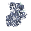
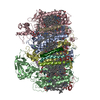
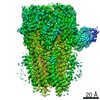
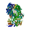
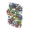
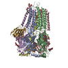
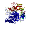
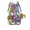
| Title | Structure of Rabies SAD-B19 L-P complex from cryo-EM | ||||||
 Components Components |
| ||||||
 Keywords Keywords | VIRAL PROTEIN / Polymerase / Phosphoprotein / Complex | ||||||
| Function / homology |  Function and homology information Function and homology informationsymbiont-mediated suppression of host transcription / GDP polyribonucleotidyltransferase / negative stranded viral RNA replication / microtubule-dependent intracellular transport of viral material towards nucleus / viral transcription / symbiont-mediated suppression of host JAK-STAT cascade via inhibition of STAT2 activity / Hydrolases; Acting on acid anhydrides; In phosphorus-containing anhydrides / symbiont-mediated suppression of host JAK-STAT cascade via inhibition of STAT1 activity / virion component / host cell ...symbiont-mediated suppression of host transcription / GDP polyribonucleotidyltransferase / negative stranded viral RNA replication / microtubule-dependent intracellular transport of viral material towards nucleus / viral transcription / symbiont-mediated suppression of host JAK-STAT cascade via inhibition of STAT2 activity / Hydrolases; Acting on acid anhydrides; In phosphorus-containing anhydrides / symbiont-mediated suppression of host JAK-STAT cascade via inhibition of STAT1 activity / virion component / host cell / symbiont-mediated suppression of host cytoplasmic pattern recognition receptor signaling pathway via inhibition of TBK1 activity / symbiont-mediated suppression of host toll-like receptor signaling pathway / host cell cytoplasm / mRNA 5'-cap (guanine-N7-)-methyltransferase activity / symbiont-mediated suppression of host type I interferon-mediated signaling pathway / RNA-directed RNA polymerase / RNA-directed RNA polymerase activity / GTPase activity / symbiont entry into host cell / host cell nucleus / ATP binding / metal ion binding Similarity search - Function | ||||||
| Biological species |  Rabies virus Rabies virus | ||||||
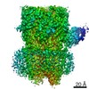
| Method | ELECTRON MICROSCOPY / single particle reconstruction / cryo EM / Resolution: 3.3 Å | ||||||
 Authors Authors | Horwitz, J.A. / Harrison, S.C. | ||||||
 Citation Citation | Journal: Proc Natl Acad Sci U S A / Year: 2020 Title: Structure of a rabies virus polymerase complex from electron cryo-microscopy. Authors: Joshua A Horwitz / Simon Jenni / Stephen C Harrison / Sean P J Whelan /  Abstract: Nonsegmented negative-stranded (NNS) RNA viruses, among them the virus that causes rabies (RABV), include many deadly human pathogens. The large polymerase (L) proteins of NNS RNA viruses carry all ...Nonsegmented negative-stranded (NNS) RNA viruses, among them the virus that causes rabies (RABV), include many deadly human pathogens. The large polymerase (L) proteins of NNS RNA viruses carry all of the enzymatic functions required for viral messenger RNA (mRNA) transcription and replication: RNA polymerization, mRNA capping, and cap methylation. We describe here a complete structure of RABV L bound with its phosphoprotein cofactor (P), determined by electron cryo-microscopy at 3.3 Å resolution. The complex closely resembles the vesicular stomatitis virus (VSV) L-P, the one other known full-length NNS-RNA L-protein structure, with key local differences (e.g., in L-P interactions). Like the VSV L-P structure, the RABV complex analyzed here represents a preinitiation conformation. Comparison with the likely elongation state, seen in two structures of pneumovirus L-P complexes, suggests differences between priming/initiation and elongation complexes. Analysis of internal cavities within RABV L suggests distinct template and product entry and exit pathways during transcription and replication. | ||||||
| History |
|
- Structure visualization
| Movie |
Movie viewer |
|---|---|
| Structure viewer | Molecule: MolmilJmol/JSmol |
- Downloads & links
-Download
| PDBx/mmCIF format | 6ueb.cif.gz | 712.4 KB | Display | PDBx/mmCIF format |
|---|---|---|---|---|
| PDB format | pdb6ueb.ent.gz | 590.4 KB | Display | PDB format |
| PDBx/mmJSON format | 6ueb.json.gz | Tree view | PDBx/mmJSON format | |
| Others |  Other downloads Other downloads |
-Validation report
| Arichive directory | https://data.pdbj.org/pub/pdb/validation_reports/ue/6uebftp://data.pdbj.org/pub/pdb/validation_reports/ue/6ueb | HTTPS FTP |
|---|
-Related structure data
| Related structure data | 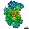 20753MC M: map data used to model this data C: citing same article ( |
|---|---|
| Similar structure data |
-Links
 PDBj
PDBj
- Assembly
| Deposited unit | 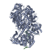
|
|---|---|
| 1 |
|
-Components
| #1: Protein | Mass: 243274.344 Da / Num. of mol.: 1 Source method: isolated from a genetically manipulated source Source: (gene. exp.) Rabies virus (strain SAD B19) / Strain: SAD B19 / Cell line (production host): SF-9 / Production host:   Spodoptera frugiperda (fall armyworm) Spodoptera frugiperda (fall armyworm)References: UniProt: P16289, RNA-directed RNA polymerase, mRNA (guanine-N7)-methyltransferase, GDP polyribonucleotidyltransferase, EC: 2.1.1.296 | ||
|---|---|---|---|
| #2: Protein/peptide | Mass: 4623.038 Da / Num. of mol.: 1 Source method: isolated from a genetically manipulated source Source: (gene. exp.) Rabies virus (strain SAD B19) / Strain: SAD B19 / Cell line (production host): SF-9 / Production host: Spodoptera frugiperda (fall armyworm) / References: UniProt: P16286 | ||
| #3: Chemical |   Mass: 65.409 Da / Num. of mol.: 2 / Source method: obtained synthetically / Formula: Zn Mass: 65.409 Da / Num. of mol.: 2 / Source method: obtained synthetically / Formula: ZnHas ligand of interest | N | |
-Experimental details
-Experiment
| Experiment | Method: ELECTRON MICROSCOPY |
|---|---|
| EM experiment | Aggregation state: PARTICLE / 3D reconstruction method: single particle reconstruction |
- Sample preparation
| Component | Name: Rabies lyssavirus strain SAD-B19 L-P complex / Type: COMPLEX Details: Purified L-P(1-91) complexes from baculovirus co-expression in insect cells Entity ID: #1-#2 / Source: RECOMBINANT |
|---|---|
| Molecular weight | Experimental value: NO |
| Source (natural) | Organism: Rabies virus (strain SAD B19) |
| Source (recombinant) | Organism: Spodoptera frugiperda (fall armyworm) / Strain: SF-9 |
| Buffer solution | pH: 8.5 |
| Specimen | Conc.: 0.3 mg/ml / Embedding applied: NO / Shadowing applied: NO / Staining applied: NO / Vitrification applied: YES / Details: This sample was monodisperse. |
| Specimen support | Details: unspecified |
| Vitrification | Cryogen name: ETHANE |
- Electron microscopy imaging
| Experimental equipment |  Model: Tecnai Polara / Image courtesy: FEI Company |
|---|---|
| Microscopy | Model: FEI POLARA 300 |
| Electron gun | Electron source:  FIELD EMISSION GUN / Accelerating voltage: 300 kV / Illumination mode: FLOOD BEAM FIELD EMISSION GUN / Accelerating voltage: 300 kV / Illumination mode: FLOOD BEAM |
| Electron lens | Mode: BRIGHT FIELD |
| Image recording | Electron dose: 72 e/Å2 / Film or detector model: GATAN K2 SUMMIT (4k x 4k) |
- Processing
| CTF correction | Type: PHASE FLIPPING AND AMPLITUDE CORRECTION |
|---|---|
| Symmetry | Point symmetry: C1 (asymmetric) |
| 3D reconstruction | Resolution: 3.3 Å / Resolution method: FSC 0.143 CUT-OFF / Num. of particles: 44500 Details: cisTEM was used for the final reconstruction and FSC curve/resolution analysis. Symmetry type: POINT |
