 Movie
Movie Controller


+ Open data

- Basic information
| Entry | Database: PDB / ID: 6tud | ||||||
|---|---|---|---|---|---|---|---|
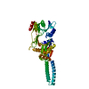
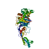
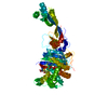
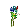
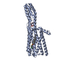
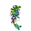
| Title | Crystal structure of Y. pestis penicillin-binding protein 3 | ||||||
 Components Components | Peptidoglycan D,D-transpeptidase FtsI | ||||||
 Keywords Keywords | CYTOSOLIC PROTEIN / Class B PBP / Yersinia pestis / HMM transpeptidase / periplasmic protein | ||||||
| Function / homology |  Function and homology information Function and homology informationpeptidoglycan glycosyltransferase activity / serine-type D-Ala-D-Ala carboxypeptidase / serine-type D-Ala-D-Ala carboxypeptidase activity / division septum assembly / FtsZ-dependent cytokinesis / penicillin binding / peptidoglycan biosynthetic process / cell wall organization / regulation of cell shape / proteolysis / plasma membrane Similarity search - Function | ||||||
| Biological species |   Yersinia pestis (bacteria) Yersinia pestis (bacteria) | ||||||
| Method |  X-RAY DIFFRACTION / SYNCHROTRON / MOLECULAR REPLACEMENT / Resolution: 3 Å X-RAY DIFFRACTION / SYNCHROTRON / MOLECULAR REPLACEMENT / Resolution: 3 Å | ||||||
 Authors Authors | Pankov, G. / Hunter, W.N. / Dawson, A. | ||||||
 Citation Citation | Journal: To Be Published Title: The structure of penicillin-binding protein 3 from Yersinia pestis Authors: Pankov, G. | ||||||
| History |
|
- Structure visualization
| Structure viewer | Molecule: MolmilJmol/JSmol |
|---|
- Downloads & links
-Download
| PDBx/mmCIF format | 6tud.cif.gz | 191.7 KB | Display | PDBx/mmCIF format |
|---|---|---|---|---|
| PDB format | pdb6tud.ent.gz | Display | PDB format | |
| PDBx/mmJSON format | 6tud.json.gz | Tree view | PDBx/mmJSON format | |
| Others |  Other downloads Other downloads |
-Validation report
| Arichive directory | https://data.pdbj.org/pub/pdb/validation_reports/tu/6tudftp://data.pdbj.org/pub/pdb/validation_reports/tu/6tud | HTTPS FTP |
|---|
-Related structure data
| Related structure data | 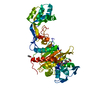 6synSC S: Starting model for refinement C: citing same article ( |
|---|---|
| Similar structure data |


-Links
 PDBj
PDBj
- Assembly
| Deposited unit | 
| ||||||||
|---|---|---|---|---|---|---|---|---|---|
| 1 | 
| ||||||||
| 2 | 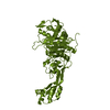
| ||||||||
| Unit cell |
|
-Components
| #1: Protein | Mass: 59558.836 Da / Num. of mol.: 2 Source method: isolated from a genetically manipulated source Source: (gene. exp.) Yersinia pestis (bacteria) / Gene: ftsI, YPO0549 / Production host: References: UniProt: Q0WJB8, serine-type D-Ala-D-Ala carboxypeptidase #2: Water | ChemComp-HOH / |  Mass: 18.015 Da / Num. of mol.: 27 / Source method: isolated from a natural source / Formula: H2O Mass: 18.015 Da / Num. of mol.: 27 / Source method: isolated from a natural source / Formula: H2O |
|---|
-Experimental details
-Experiment
| Experiment | Method: X-RAY DIFFRACTION / Number of used crystals: 1 |
|---|
- Sample preparation
| Crystal | Density Matthews: 3.47 Å3/Da / Density % sol: 65 % |
|---|---|
| Crystal grow | Temperature: 296 K / Method: vapor diffusion, sitting drop Details: 1 uL of protein (in 20 mM Tris-HCl pH 7.5 150 mM NaCl and 2 mM carbenicillin) at 7 mg/ml and 0.2 uL of precipitant (0.2 M magnesium acetate, 6% gamma-PGA (Na+ form, LM) and 6% PEG8000) ...Details: 1 uL of protein (in 20 mM Tris-HCl pH 7.5 150 mM NaCl and 2 mM carbenicillin) at 7 mg/ml and 0.2 uL of precipitant (0.2 M magnesium acetate, 6% gamma-PGA (Na+ form, LM) and 6% PEG8000) Crystals were then dehydrated by replacing 50% of the reservoir solution with 50% PEG10000 |
-Data collection
| Diffraction | Mean temperature: 100 K / Serial crystal experiment: N |
|---|---|
| Diffraction source | Source: SYNCHROTRON / Site: Diamond  / Beamline: I04-1 / Wavelength: 0.9196 Å / Beamline: I04-1 / Wavelength: 0.9196 Å |
| Detector | Type: DECTRIS PILATUS 6M-F / Detector: PIXEL / Date: Jan 17, 2018 |
| Radiation | Protocol: SINGLE WAVELENGTH / Monochromatic (M) / Laue (L): M / Scattering type: x-ray |
| Radiation wavelength | Wavelength: 0.9196 Å / Relative weight: 1 |
| Reflection | Resolution: 3→29.64 Å / Num. obs: 33546 / % possible obs: 99.9 % / Redundancy: 10 % / Biso Wilson estimate: 50.6 Å2 / CC1/2: 0.997 / Rmerge(I) obs: 0.2 / Rpim(I) all: 0.071 / Net I/σ(I): 9.1 |
| Reflection shell | Resolution: 3→3.15 Å / Rmerge(I) obs: 1.099 / Mean I/σ(I) obs: 1.4 / Num. unique obs: 4365 / CC1/2: 0.941 / Rpim(I) all: 0.518 / % possible all: 100 |
- Processing
| Software |
| ||||||||||||||||||||||||||||||||||||||||||||||||||||||||||||||||||||||||||||||||||||||||||||||||||||||||||||||||||||||||||||||||||||||||||||||||||||||
|---|---|---|---|---|---|---|---|---|---|---|---|---|---|---|---|---|---|---|---|---|---|---|---|---|---|---|---|---|---|---|---|---|---|---|---|---|---|---|---|---|---|---|---|---|---|---|---|---|---|---|---|---|---|---|---|---|---|---|---|---|---|---|---|---|---|---|---|---|---|---|---|---|---|---|---|---|---|---|---|---|---|---|---|---|---|---|---|---|---|---|---|---|---|---|---|---|---|---|---|---|---|---|---|---|---|---|---|---|---|---|---|---|---|---|---|---|---|---|---|---|---|---|---|---|---|---|---|---|---|---|---|---|---|---|---|---|---|---|---|---|---|---|---|---|---|---|---|---|---|---|---|
| Refinement | Method to determine structure: MOLECULAR REPLACEMENT Starting model: 6SYN Resolution: 3→29.64 Å / Cor.coef. Fo:Fc: 0.935 / Cor.coef. Fo:Fc free: 0.91 / SU B: 26.209 / SU ML: 0.403 / Cross valid method: FREE R-VALUE / ESU R: 1.048 / ESU R Free: 0.398 Details: Hydrogens have been added in their riding positions
| ||||||||||||||||||||||||||||||||||||||||||||||||||||||||||||||||||||||||||||||||||||||||||||||||||||||||||||||||||||||||||||||||||||||||||||||||||||||
| Solvent computation | Ion probe radii: 0.8 Å / Shrinkage radii: 0.8 Å / VDW probe radii: 1.2 Å | ||||||||||||||||||||||||||||||||||||||||||||||||||||||||||||||||||||||||||||||||||||||||||||||||||||||||||||||||||||||||||||||||||||||||||||||||||||||
| Displacement parameters | Biso mean: 82.519 Å2
| ||||||||||||||||||||||||||||||||||||||||||||||||||||||||||||||||||||||||||||||||||||||||||||||||||||||||||||||||||||||||||||||||||||||||||||||||||||||
| Refinement step | Cycle: LAST / Resolution: 3→29.64 Å
| ||||||||||||||||||||||||||||||||||||||||||||||||||||||||||||||||||||||||||||||||||||||||||||||||||||||||||||||||||||||||||||||||||||||||||||||||||||||
| Refine LS restraints |
| ||||||||||||||||||||||||||||||||||||||||||||||||||||||||||||||||||||||||||||||||||||||||||||||||||||||||||||||||||||||||||||||||||||||||||||||||||||||
| LS refinement shell |
|
