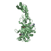
Entry Database : PDB / ID : 6snrTitle Crystal structure of FemX Lipid II:glycine glycyltransferase Keywords / / / / / / Function / homology Function Domain/homology Component
/ / / / / Biological species Staphylococcus aureus (bacteria)Method / / / Resolution : 1.62 Å Authors Fulop, V. / Hinxman, K. Funding support Organization Grant number Country Medical Research Council (United Kingdom) MR/M017893/1 Medical Research Council (United Kingdom) MR/N002679/1 Biotechnology and Biological Sciences Research Council Studentship
Journal : Structure / Year : 2021Title : Structure-based modeling and dynamics of MurM, a Streptococcus pneumoniae penicillin resistance determinant present at the cytoplasmic membrane.Authors : York, A. / Lloyd, A.J. / Del Genio, C.I. / Shearer, J. / Hinxman, K.J. / Fritz, K. / Fulop, V. / Dowson, C.G. / Khalid, S. / Roper, D.I. History Deposition Aug 27, 2019 Deposition site / Processing site Revision 1.0 Sep 9, 2020 Provider / Type Revision 1.1 Mar 23, 2022 Group / Category / citation_author / database_2Item _citation.country / _citation.journal_abbrev ... _citation.country / _citation.journal_abbrev / _citation.journal_id_ASTM / _citation.journal_id_CSD / _citation.journal_id_ISSN / _citation.journal_volume / _citation.page_first / _citation.page_last / _citation.pdbx_database_id_DOI / _citation.pdbx_database_id_PubMed / _citation.title / _citation.year / _database_2.pdbx_DOI / _database_2.pdbx_database_accession Revision 1.2 Jun 19, 2024 Group / Category / chem_comp_bond
Show all Show less
 Movie
Movie Controller


 Open data
 Basic information
 Components
 Keywords
 Function and homology information
 Staphylococcus aureus (bacteria)
 X-RAY DIFFRACTION /
  Authors
 United Kingdom, 3items
  Citation
 Structure visualization
 Downloads & links
 Other downloads


 PDBj
PDBj

 Assembly
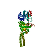
 Mass: 18.015 Da / Num. of mol.: 177 / Source method: isolated from a natural source / Formula: H2O
 Sample preparation
 Processing