 Movie
Movie Controller

[English] 日本語
 Yorodumi
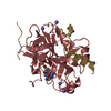
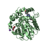
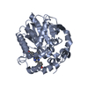
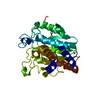
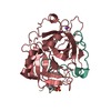
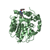
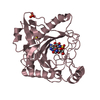
- PDB-6tkg: Tsetse thrombin inhibitor in complex with human alpha-thrombin - ... -
+ Open data

- Basic information
| Entry | Database: PDB / ID: 6tkg | |||||||||
|---|---|---|---|---|---|---|---|---|---|---|
| Title | Tsetse thrombin inhibitor in complex with human alpha-thrombin - orthorhombic form at 12keV | |||||||||
 Components Components |
| |||||||||
 Keywords Keywords | BLOOD CLOTTING / anticoagulant tyrosine sulfation posttranslational modification complex | |||||||||
| Function / homology |  Function and homology information Function and homology informationcytolysis by host of symbiont cells / thrombospondin receptor activity / thrombin-activated receptor signaling pathway / Defective factor XII causes hereditary angioedema / thrombin / negative regulation of astrocyte differentiation / regulation of blood coagulation / positive regulation of phospholipase C-activating G protein-coupled receptor signaling pathway / neutrophil-mediated killing of gram-negative bacterium / Defective F8 cleavage by thrombin ...cytolysis by host of symbiont cells / thrombospondin receptor activity / thrombin-activated receptor signaling pathway / Defective factor XII causes hereditary angioedema / thrombin / negative regulation of astrocyte differentiation / regulation of blood coagulation / positive regulation of phospholipase C-activating G protein-coupled receptor signaling pathway / neutrophil-mediated killing of gram-negative bacterium / Defective F8 cleavage by thrombin / Platelet Aggregation (Plug Formation) / ligand-gated ion channel signaling pathway / positive regulation of collagen biosynthetic process / negative regulation of platelet activation / negative regulation of blood coagulation / positive regulation of blood coagulation / negative regulation of fibrinolysis / regulation of cytosolic calcium ion concentration / Transport of gamma-carboxylated protein precursors from the endoplasmic reticulum to the Golgi apparatus / Gamma-carboxylation of protein precursors / Common Pathway of Fibrin Clot Formation / Removal of aminoterminal propeptides from gamma-carboxylated proteins / fibrinolysis / Intrinsic Pathway of Fibrin Clot Formation / negative regulation of proteolysis / negative regulation of cytokine production involved in inflammatory response / positive regulation of release of sequestered calcium ion into cytosol / Peptide ligand-binding receptors / Regulation of Complement cascade / acute-phase response / Cell surface interactions at the vascular wall / positive regulation of receptor signaling pathway via JAK-STAT / lipopolysaccharide binding / growth factor activity / serine-type endopeptidase inhibitor activity / positive regulation of insulin secretion / platelet activation / response to wounding / positive regulation of protein localization to nucleus / Golgi lumen / Regulation of Insulin-like Growth Factor (IGF) transport and uptake by Insulin-like Growth Factor Binding Proteins (IGFBPs) / positive regulation of reactive oxygen species metabolic process / antimicrobial humoral immune response mediated by antimicrobial peptide / blood coagulation / regulation of cell shape / heparin binding / Thrombin signalling through proteinase activated receptors (PARs) / : / positive regulation of cell growth / blood microparticle / G alpha (q) signalling events / cell surface receptor signaling pathway / positive regulation of phosphatidylinositol 3-kinase/protein kinase B signal transduction / receptor ligand activity / endoplasmic reticulum lumen / signaling receptor binding / serine-type endopeptidase activity / positive regulation of cell population proliferation / calcium ion binding / proteolysis / extracellular space / extracellular exosome / extracellular region / plasma membrane Similarity search - Function | |||||||||
| Biological species |  Homo sapiens (human) Homo sapiens (human) Glossina morsitans morsitans (fry) Glossina morsitans morsitans (fry) | |||||||||
| Method |  X-RAY DIFFRACTION / SYNCHROTRON / MOLECULAR REPLACEMENT / Resolution: 1.35 Å X-RAY DIFFRACTION / SYNCHROTRON / MOLECULAR REPLACEMENT / Resolution: 1.35 Å | |||||||||
 Authors Authors | Calisto, B.M. / Ripoll-Rozada, J. / de Sanctis, D. / Pereira, P.J.B. | |||||||||
| Funding support |  Australia, Australia,  Portugal, 2items Portugal, 2items
| |||||||||
 Citation Citation | Journal: Cell Chem Biol / Year: 2021 Title: Sulfotyrosine-Mediated Recognition of Human Thrombin by a Tsetse Fly Anticoagulant Mimics Physiological Substrates. Authors: Calisto, B.M. / Ripoll-Rozada, J. / Dowman, L.J. / Franck, C. / Agten, S.M. / Parker, B.L. / Veloso, R.C. / Vale, N. / Gomes, P. / de Sanctis, D. / Payne, R.J. / Pereira, P.J.B. | |||||||||
| History |
|
- Structure visualization
| Structure viewer | Molecule: MolmilJmol/JSmol |
|---|
- Downloads & links
-Download
| PDBx/mmCIF format | 6tkg.cif.gz | 172.6 KB | Display | PDBx/mmCIF format |
|---|---|---|---|---|
| PDB format | pdb6tkg.ent.gz | 116.8 KB | Display | PDB format |
| PDBx/mmJSON format | 6tkg.json.gz | Tree view | PDBx/mmJSON format | |
| Others |  Other downloads Other downloads |
-Validation report
| Summary document | 6tkg_validation.pdf.gz | 477.7 KB | Display | wwPDB validaton report |
|---|---|---|---|---|
| Full document | 6tkg_full_validation.pdf.gz | 479.6 KB | Display | |
| Data in XML | 6tkg_validation.xml.gz | 17.6 KB | Display | |
| Data in CIF | 6tkg_validation.cif.gz | 25.8 KB | Display | |
| Arichive directory | https://data.pdbj.org/pub/pdb/validation_reports/tk/6tkgftp://data.pdbj.org/pub/pdb/validation_reports/tk/6tkg | HTTPS FTP |
-Related structure data
| Related structure data | 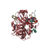 6tkhC 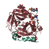 6tkiC 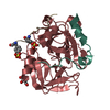 6tkjC 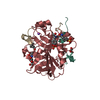 6tklC  3u69S S: Starting model for refinement C: citing same article ( |
|---|---|
| Similar structure data | |
| Experimental dataset #1 | Data reference: 10.15785/SBGRID/737 / Data set type: diffraction image data |


-Links
 PDBj
PDBj


- Assembly
| Deposited unit | 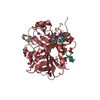
| ||||||||||||
|---|---|---|---|---|---|---|---|---|---|---|---|---|---|
| 1 |
| ||||||||||||
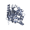
| Unit cell |
|
-Components
-Protein , 2 types, 2 molecules HI
| #2: Protein | Mass: 29780.219 Da / Num. of mol.: 1 / Source method: isolated from a natural source / Source: (natural) Homo sapiens (human) / References: UniProt: P00734, thrombin |
|---|---|
| #3: Protein | Mass: 5962.750 Da / Num. of mol.: 1 / Source method: obtained synthetically Details: Residues 9 and 12 of the mature sequence are O-sulfated. Source: (synth.) Glossina morsitans morsitans (fry) / References: UniProt: O97373 |
-Protein/peptide / Sugars , 2 types, 2 molecules L
| #1: Protein/peptide | Mass: 4096.534 Da / Num. of mol.: 1 / Source method: isolated from a natural source / Source: (natural) Homo sapiens (human) / References: UniProt: P00734, thrombin |
|---|---|
| #4: Sugar | ChemComp-NAG /  Type: D-saccharide, beta linking / Mass: 221.208 Da / Num. of mol.: 1 Type: D-saccharide, beta linking / Mass: 221.208 Da / Num. of mol.: 1Source method: isolated from a genetically manipulated source Formula: C8H15NO6 |
-Non-polymers , 3 types, 312 molecules 


| #5: Chemical | ChemComp-GOL /  Mass: 92.094 Da / Num. of mol.: 1 / Source method: obtained synthetically / Formula: C3H8O3 Mass: 92.094 Da / Num. of mol.: 1 / Source method: obtained synthetically / Formula: C3H8O3 |
|---|---|
| #6: Chemical | ChemComp-NA /  Mass: 22.990 Da / Num. of mol.: 1 / Source method: obtained synthetically / Formula: Na Mass: 22.990 Da / Num. of mol.: 1 / Source method: obtained synthetically / Formula: Na |
| #7: Water | ChemComp-HOH / Mass: 18.015 Da / Num. of mol.: 310 / Source method: isolated from a natural source / Formula: H2O |
-Details
| Has ligand of interest | Y |
|---|---|
| Has protein modification | Y |
-Experimental details
-Experiment
| Experiment | Method: X-RAY DIFFRACTION / Number of used crystals: 1 |
|---|
- Sample preparation
| Crystal | Density Matthews: 2.23 Å3/Da / Density % sol: 44.8 % |
|---|---|
| Crystal grow | Temperature: 293 K / Method: vapor diffusion, sitting drop Details: 0.1 M HEPES pH 7.5, 0.2 M ammonium acetate, 25 % (w/v) PEG 3350 |
-Data collection
| Diffraction | Mean temperature: 100 K / Serial crystal experiment: N |
|---|---|
| Diffraction source | Source: SYNCHROTRON / Site: ALBA  / Beamline: XALOC / Wavelength: 1.033 Å / Beamline: XALOC / Wavelength: 1.033 Å |
| Detector | Type: DECTRIS PILATUS 6M / Detector: PIXEL / Date: Oct 20, 2017 |
| Radiation | Protocol: SINGLE WAVELENGTH / Monochromatic (M) / Laue (L): M / Scattering type: x-ray |
| Radiation wavelength | Wavelength: 1.033 Å / Relative weight: 1 |
| Reflection | Resolution: 1.35→58.9 Å / Num. obs: 70747 / % possible obs: 98.5 % / Redundancy: 5.4 % / Biso Wilson estimate: 17.3 Å2 / CC1/2: 0.999 / Rmerge(I) obs: 0.073 / Net I/σ(I): 9.7 |
| Reflection shell | Resolution: 1.35→1.37 Å / Redundancy: 5 % / Rmerge(I) obs: 2.34 / Mean I/σ(I) obs: 0.5 / Num. unique obs: 3266 / CC1/2: 0.421 / % possible all: 91.3 |
- Processing
| Software |
| |||||||||||||||||||||||||||||||||||||||||||||||||||||||||||||||||||||||||||||||||||||||||||||||||||||||||||||||||||||||||||||||||||||||||||||||||||||||||||||||||||||||||||||||
|---|---|---|---|---|---|---|---|---|---|---|---|---|---|---|---|---|---|---|---|---|---|---|---|---|---|---|---|---|---|---|---|---|---|---|---|---|---|---|---|---|---|---|---|---|---|---|---|---|---|---|---|---|---|---|---|---|---|---|---|---|---|---|---|---|---|---|---|---|---|---|---|---|---|---|---|---|---|---|---|---|---|---|---|---|---|---|---|---|---|---|---|---|---|---|---|---|---|---|---|---|---|---|---|---|---|---|---|---|---|---|---|---|---|---|---|---|---|---|---|---|---|---|---|---|---|---|---|---|---|---|---|---|---|---|---|---|---|---|---|---|---|---|---|---|---|---|---|---|---|---|---|---|---|---|---|---|---|---|---|---|---|---|---|---|---|---|---|---|---|---|---|---|---|---|---|---|
| Refinement | Method to determine structure: MOLECULAR REPLACEMENT Starting model: 3U69 Resolution: 1.35→42.77 Å / SU ML: 0.2059 / Cross valid method: FREE R-VALUE / σ(F): 1.33 / Phase error: 25.2386
| |||||||||||||||||||||||||||||||||||||||||||||||||||||||||||||||||||||||||||||||||||||||||||||||||||||||||||||||||||||||||||||||||||||||||||||||||||||||||||||||||||||||||||||||
| Solvent computation | Shrinkage radii: 0.9 Å / VDW probe radii: 1.11 Å | |||||||||||||||||||||||||||||||||||||||||||||||||||||||||||||||||||||||||||||||||||||||||||||||||||||||||||||||||||||||||||||||||||||||||||||||||||||||||||||||||||||||||||||||
| Displacement parameters | Biso mean: 27.88 Å2 | |||||||||||||||||||||||||||||||||||||||||||||||||||||||||||||||||||||||||||||||||||||||||||||||||||||||||||||||||||||||||||||||||||||||||||||||||||||||||||||||||||||||||||||||
| Refinement step | Cycle: LAST / Resolution: 1.35→42.77 Å
| |||||||||||||||||||||||||||||||||||||||||||||||||||||||||||||||||||||||||||||||||||||||||||||||||||||||||||||||||||||||||||||||||||||||||||||||||||||||||||||||||||||||||||||||
| Refine LS restraints |
| |||||||||||||||||||||||||||||||||||||||||||||||||||||||||||||||||||||||||||||||||||||||||||||||||||||||||||||||||||||||||||||||||||||||||||||||||||||||||||||||||||||||||||||||
| LS refinement shell |
|
