 Movie
Movie Controller


+ Open data

- Basic information
| Entry | Database: PDB / ID: 6tbu | |||||||||||||||||||||||||||||||||||||||
|---|---|---|---|---|---|---|---|---|---|---|---|---|---|---|---|---|---|---|---|---|---|---|---|---|---|---|---|---|---|---|---|---|---|---|---|---|---|---|---|---|
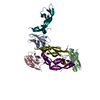
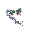
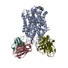
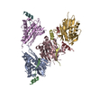
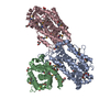
| Title | Structure of Drosophila melanogaster Dispatched | |||||||||||||||||||||||||||||||||||||||
 Components Components | Protein dispatched | |||||||||||||||||||||||||||||||||||||||
 Keywords Keywords | MEMBRANE PROTEIN / RND transporter / transmembrane domain / ectodomain / cholesteryl hemisuccinate / detergent micelle / digitonin / monomer | |||||||||||||||||||||||||||||||||||||||
| Function / homology |  Function and homology information Function and homology informationFormation and transport of the N-HH ligand / cytoneme / wing disc pattern formation / patched ligand maturation / segment polarity determination / primordial germ cell migration / smoothened signaling pathway / membrane / plasma membrane Similarity search - Function | |||||||||||||||||||||||||||||||||||||||
| Biological species |  | |||||||||||||||||||||||||||||||||||||||
| Method | ELECTRON MICROSCOPY / single particle reconstruction / cryo EM / Resolution: 3.16 Å | |||||||||||||||||||||||||||||||||||||||
 Authors Authors | Korkhov, V.M. / Cannac, F. | |||||||||||||||||||||||||||||||||||||||
| Funding support |  Switzerland, 1items Switzerland, 1items
| |||||||||||||||||||||||||||||||||||||||
 Citation Citation | Journal: Sci Adv / Year: 2020 Title: Cryo-EM structure of the Hedgehog release protein Dispatched. Authors: Fabien Cannac / Chao Qi / Julia Falschlunger / George Hausmann / Konrad Basler / Volodymyr M Korkhov / Abstract: The Hedgehog (Hh) signaling pathway controls embryonic development and adult tissue homeostasis in multicellular organisms. In , the pathway is primed by secretion of a dually lipid-modified ...The Hedgehog (Hh) signaling pathway controls embryonic development and adult tissue homeostasis in multicellular organisms. In , the pathway is primed by secretion of a dually lipid-modified morphogen, Hh, a process dependent on a membrane-integral protein Dispatched. Although Dispatched is a critical component of the pathway, the structural basis of its activity has, so far, not been described. Here, we describe a cryo-electron microscopy structure of the Dispatched at 3.2-Å resolution. The ectodomains of Dispatched adopt an open conformation suggestive of a receptor-chaperone role. A three-dimensional reconstruction of Dispatched bound to Hh confirms the ability of Dispatched to bind Hh but using a unique mode distinct from those previously observed in structures of Hh complexes. The structure may represent the state of the complex that precedes shedding of Hh from the surface of the morphogen-releasing cell. | |||||||||||||||||||||||||||||||||||||||
| History |
|
- Structure visualization
| Movie |
Movie viewer |
|---|---|
| Structure viewer | Molecule: MolmilJmol/JSmol |
- Downloads & links
-Download
| PDBx/mmCIF format | 6tbu.cif.gz | 311.3 KB | Display | PDBx/mmCIF format |
|---|---|---|---|---|
| PDB format | pdb6tbu.ent.gz | 252.2 KB | Display | PDB format |
| PDBx/mmJSON format | 6tbu.json.gz | Tree view | PDBx/mmJSON format | |
| Others |  Other downloads Other downloads |
-Validation report
| Arichive directory | https://data.pdbj.org/pub/pdb/validation_reports/tb/6tbuftp://data.pdbj.org/pub/pdb/validation_reports/tb/6tbu | HTTPS FTP |
|---|
-Related structure data
| Related structure data |  10452MC  6td6C M: map data used to model this data C: citing same article ( |
|---|---|
| Similar structure data |


-Links
 PDBj
PDBj
- Assembly
| Deposited unit | 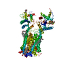
|
|---|---|
| 1 |
|
-Components
| #1: Protein | Mass: 139149.875 Da / Num. of mol.: 1 Source method: isolated from a genetically manipulated source Details: Drosophila melanogaster Dispatched, tagged with a C-terminal 3C-protease cleavage site, YFP and twin-strep tag. Source: (gene. exp.)  Homo sapiens (human) / References: UniProt: Q9VNJ5 Homo sapiens (human) / References: UniProt: Q9VNJ5 | ||||||
|---|---|---|---|---|---|---|---|
| #2: Polysaccharide | 2-acetamido-2-deoxy-beta-D-glucopyranose-(1-4)-2-acetamido-2-deoxy-beta-D-glucopyranose Source method: isolated from a genetically manipulated source | ||||||
| #3: Chemical | ChemComp-Y01 /   Mass: 486.726 Da / Num. of mol.: 9 / Source method: obtained synthetically / Formula: C31H50O4 Mass: 486.726 Da / Num. of mol.: 9 / Source method: obtained synthetically / Formula: C31H50O4#4: Sugar | ChemComp-NAG / |   Type: D-saccharide, beta linking / Mass: 221.208 Da / Num. of mol.: 1 Type: D-saccharide, beta linking / Mass: 221.208 Da / Num. of mol.: 1Source method: isolated from a genetically manipulated source Formula: C8H15NO6 Has ligand of interest | N | Has protein modification | Y | |
-Experimental details
-Experiment
| Experiment | Method: ELECTRON MICROSCOPY |
|---|---|
| EM experiment | Aggregation state: PARTICLE / 3D reconstruction method: single particle reconstruction |
- Sample preparation
| Component | Name: Drosophila melanogaster protein Dispatched (disp) / Type: COMPLEX / Entity ID: #1 / Source: RECOMBINANT |
|---|---|
| Molecular weight | Value: 0.139 MDa / Experimental value: NO |
| Source (natural) | Organism: |
| Source (recombinant) | Organism: Homo sapiens (human) |
| Buffer solution | pH: 8 |
| Specimen | Embedding applied: NO / Shadowing applied: NO / Staining applied: NO / Vitrification applied: YES |
| Vitrification | Cryogen name: ETHANE |
- Electron microscopy imaging
| Experimental equipment |  Model: Titan Krios / Image courtesy: FEI Company |
|---|---|
| Microscopy | Model: FEI TITAN KRIOS |
| Electron gun | Electron source:  FIELD EMISSION GUN / Accelerating voltage: 300 kV / Illumination mode: FLOOD BEAM FIELD EMISSION GUN / Accelerating voltage: 300 kV / Illumination mode: FLOOD BEAM |
| Electron lens | Mode: BRIGHT FIELD / Nominal defocus max: 2400 nm / Nominal defocus min: 800 nm |
| Image recording | Electron dose: 50 e/Å2 / Detector mode: COUNTING / Film or detector model: GATAN K2 QUANTUM (4k x 4k) / Num. of real images: 9216 |
- Processing
| EM software |
| ||||||||||||||||
|---|---|---|---|---|---|---|---|---|---|---|---|---|---|---|---|---|---|
| CTF correction | Type: PHASE FLIPPING AND AMPLITUDE CORRECTION | ||||||||||||||||
| Symmetry | Point symmetry: C1 (asymmetric) | ||||||||||||||||
| 3D reconstruction | Resolution: 3.16 Å / Resolution method: FSC 0.143 CUT-OFF / Num. of particles: 286136 / Symmetry type: POINT |
