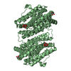
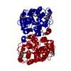
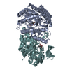
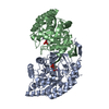
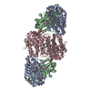
- PDB-2rcc: Crystal structure of putative class I ribonucleotide reductase (N... -
+
Open data
ID or keywords:
Loading...
-
Basic information
Entry
Database: PDB / ID: 2rcc
Title
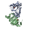
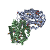
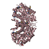
Crystal structure of putative class I ribonucleotide reductase (NP_241368.1) from Bacillus halodurans at 1.90 A resolution
Components
Ribonucleoside-diphosphate reductase subunit beta
Keywords
OXIDOREDUCTASE / NP_241368.1 / Putative Class I Ribonucleotide Reductase / Ribonucleotide reductase / small chain / Structural Genomics / Joint Center for Structural Genomics / JCSG / Protein Structure Initiative / PSI-2 / DNA replication / Iron / Metal-binding
Function / homology
Function and homology information
ribonucleoside-diphosphate reductase / ribonucleoside-diphosphate reductase activity, thioredoxin disulfide as acceptor / deoxyribonucleotide biosynthetic process / metal ion binding Similarity search - Function
Ribonucleotide Reductase, subunit A / Ribonucleotide Reductase, subunit A / Ribonucleotide reductase small subunit / Ribonucleotide reductase small subunit family / Ribonucleotide reductase, small chain / Ribonucleotide reductase-like / Ferritin-like superfamily / Orthogonal Bundle / Mainly Alpha Similarity search - Domain/homology
BIOMOLECULE: 1, 2, 3 SEE REMARK 350 FOR THE AUTHOR PROVIDED AND PROGRAM GENERATED ASSEMBLY ... BIOMOLECULE: 1, 2, 3 SEE REMARK 350 FOR THE AUTHOR PROVIDED AND PROGRAM GENERATED ASSEMBLY INFORMATION FOR THE STRUCTURE IN THIS ENTRY. THE REMARK MAY ALSO PROVIDE INFORMATION ON BURIED SURFACE AREA. THE ASSIGNMENT OF A DIMER AS THE SIGNIFICANT OLIGOMERIZATION STATE IN SOLUTION IS SUPPORTED BY SIZE EXCLUSION CHROMATOGRAPHY AND STATIC LIGHT SCATTERING.
Remark 999
SEQUENCE THE CONSTRUCT WAS EXPRESSED WITH A PURIFICATION TAG MGSDKIHHHHHHENLYFQG. THE TAG WAS ... SEQUENCE THE CONSTRUCT WAS EXPRESSED WITH A PURIFICATION TAG MGSDKIHHHHHHENLYFQG. THE TAG WAS REMOVED WITH TEV PROTEASE LEAVING ONLY A GLYCINE FOLLOWED BY THE TARGET SEQUENCE.
THE ASSIGNMENT OF A DIMER AS THE SIGNIFICANT OLIGOMERIZATION STATE IN SOLUTION IS SUPPORTED BY SIZE EXCLUSION CHROMATOGRAPHY AND STATIC LIGHT SCATTERING.
-
Components
-
Protein , 1 types, 3 molecules ABC
#1: Protein
Ribonucleoside-diphosphatereductasesubunitbeta / Ribonucleotide reductase small subunit
Mass: 40855.270 Da / Num. of mol.: 3 Source method: isolated from a genetically manipulated source Source: (gene. exp.) Bacillus halodurans C-125 (bacteria) / Species: Bacillus halodurans / Strain: C-125, DSM 18197, FERM 7344, JCM 9153 / Gene: NP_241368.1, nrdB, BH0502 / Plasmid: speedET / Production host: Escherichia coli (E. coli) / Strain (production host): HK100 References: UniProt: Q9KFH7, ribonucleoside-diphosphate reductase
Monochromator: Single crystal Si(111) bent (horizontal focusing) Protocol: MAD / Monochromatic (M) / Laue (L): M / Scattering type: x-ray
Radiation wavelength
ID
Wavelength (Å)
Relative weight
1
0.91837
1
2
0.97926
1
Reflection
Resolution: 1.9→28.502 Å / Num. obs: 90521 / % possible obs: 98.2 % / Observed criterion σ(I): -3 / Biso Wilson estimate: 27.44 Å2 / Rmerge(I) obs: 0.055 / Net I/σ(I): 7.13
Reflection shell
Resolution (Å)
Rmerge(I) obs
Mean I/σ(I) obs
Num. measured obs
Num. unique obs
Diffraction-ID
% possible all
1.9-1.97
0.389
1.7
21432
17841
1
97
1.97-2.05
0.299
2.3
21353
17649
1
98.3
2.05-2.14
0.219
3
20733
16951
1
98.3
2.14-2.25
0.165
4
21043
17259
1
98.6
2.25-2.39
0.127
5
21498
17591
1
98.8
2.39-2.58
0.097
6.4
22405
18280
1
98.9
2.58-2.84
0.07
8.3
21609
17685
1
98.9
2.84-3.25
0.054
10.7
21563
17636
1
98.8
3.25-4.08
0.041
14
20910
17470
1
98.7
4.08-28.502
0.032
16
21354
17437
1
96
-
Phasing
Phasing
Method: MAD
-
Processing
Software
Name
Version
Classification
NB
REFMAC
5.3.0040
refinement
PHENIX
refinement
SHELX
phasing
MolProbity
3beta29
modelbuilding
XSCALE
datascaling
PDB_EXTRACT
3
dataextraction
MAR345
CCD
datacollection
XDS
datareduction
SHELXD
phasing
SHARP
phasing
Refinement
Method to determine structure: MAD / Resolution: 1.9→28.502 Å / Cor.coef. Fo:Fc: 0.96 / Cor.coef. Fo:Fc free: 0.949 / SU B: 7.078 / SU ML: 0.1 / TLS residual ADP flag: LIKELY RESIDUAL / Cross valid method: THROUGHOUT / σ(F): 0 / ESU R: 0.135 / ESU R Free: 0.126 Stereochemistry target values: MAXIMUM LIKELIHOOD WITH PHASES Details: 1. HYDROGENS HAVE BEEN ADDED IN THE RIDING POSITIONS. 2. A MET-INHIBITION PROTOCOL WAS USED FOR SELENOMETHIONINE INCORPORATION DURING PROTEIN EXPRESSION. THE OCCUPANCY OF THE SE ATOMS IN THE ...Details: 1. HYDROGENS HAVE BEEN ADDED IN THE RIDING POSITIONS. 2. A MET-INHIBITION PROTOCOL WAS USED FOR SELENOMETHIONINE INCORPORATION DURING PROTEIN EXPRESSION. THE OCCUPANCY OF THE SE ATOMS IN THE MSE RESIDUES WAS REDUCED TO 0.75 TO ACCOUNT FOR THE REDUCED SCATTERING POWER DUE TO PARTIAL S-MET INCORPORATION. 3. ATOM RECORD CONTAINS RESIDUAL B FACTORS ONLY. 4. ZINC HAS BEEN MODELED IN THE PUTATIVE ACTIVE SITES OF MOLECULES B AND C BASED ON A X-RAY FLUORESCENCE SCAN FOR METAL, PRESENCE OF ELECTRON DENSITY AND COORDINATION GEOMETRY. 5. ETHYLENE GLYCOL, GLYCEROL, AND PARTIAL PEG MOLECULES HAVE BEEN MODELED IN THE SOLVENT STRUCTURE. 6. THERE IS A LARGE BLOB OF UNIDENTIFIED DENSITY IN THE VICINITY OF AMINO ACIDS A25-A28.
Rfactor
Num. reflection
% reflection
Selection details
Rfree
0.215
4538
5 %
RANDOM
Rwork
0.183
-
-
-
obs
0.184
90520
99.52 %
-
Solvent computation
Ion probe radii: 0.8 Å / Shrinkage radii: 0.8 Å / VDW probe radii: 1.2 Å / Solvent model: MASK
Displacement parameters
Biso mean: 19.585 Å2
Baniso -1
Baniso -2
Baniso -3
1-
-1.23 Å2
0 Å2
-0.95 Å2
2-
-
-0.46 Å2
0 Å2
3-
-
-
0.6 Å2
Refinement step
Cycle: LAST / Resolution: 1.9→28.502 Å
Protein
Nucleic acid
Ligand
Solvent
Total
Num. atoms
7328
0
80
340
7748
Refine LS restraints
Refine-ID
Type
Dev ideal
Dev ideal target
Number
X-RAY DIFFRACTION
r_bond_refined_d
0.017
0.022
7762
X-RAY DIFFRACTION
r_bond_other_d
0.004
0.02
5146
X-RAY DIFFRACTION
r_angle_refined_deg
1.613
1.947
10567
X-RAY DIFFRACTION
r_angle_other_deg
1.292
3
12592
X-RAY DIFFRACTION
r_dihedral_angle_1_deg
3.635
5
954
X-RAY DIFFRACTION
r_dihedral_angle_2_deg
34.801
24.84
374
X-RAY DIFFRACTION
r_dihedral_angle_3_deg
12.015
15
1286
X-RAY DIFFRACTION
r_dihedral_angle_4_deg
17.402
15
23
X-RAY DIFFRACTION
r_chiral_restr
0.092
0.2
1162
X-RAY DIFFRACTION
r_gen_planes_refined
0.007
0.02
8598
X-RAY DIFFRACTION
r_gen_planes_other
0.002
0.02
1623
X-RAY DIFFRACTION
r_nbd_refined
0.194
0.3
1811
X-RAY DIFFRACTION
r_nbd_other
0.141
0.3
5067
X-RAY DIFFRACTION
r_nbtor_refined
0.183
0.5
3861
X-RAY DIFFRACTION
r_nbtor_other
0.088
0.5
3452
X-RAY DIFFRACTION
r_xyhbond_nbd_refined
0.175
0.5
561
X-RAY DIFFRACTION
r_xyhbond_nbd_other
0.063
0.5
1
X-RAY DIFFRACTION
r_metal_ion_refined
0.071
0.5
4
X-RAY DIFFRACTION
r_symmetry_vdw_refined
0.144
0.3
11
X-RAY DIFFRACTION
r_symmetry_vdw_other
0.187
0.3
77
X-RAY DIFFRACTION
r_symmetry_hbond_refined
0.218
0.5
23
X-RAY DIFFRACTION
r_mcbond_it
2.158
3
5257
X-RAY DIFFRACTION
r_mcbond_other
0.578
3
1825
X-RAY DIFFRACTION
r_mcangle_it
2.932
5
7533
X-RAY DIFFRACTION
r_scbond_it
5.537
8
3551
X-RAY DIFFRACTION
r_scangle_it
7.089
11
3009
LS refinement shell
Resolution: 1.9→1.949 Å / Total num. of bins used: 20
Rfactor
Num. reflection
% reflection
Rfree
0.324
310
-
Rwork
0.255
6275
-
all
-
6585
-
obs
-
-
98.74 %
Refinement TLS params.
Method: refined / Refine-ID: X-RAY DIFFRACTION
ID
L11 (°2)
L12 (°2)
L13 (°2)
L22 (°2)
L23 (°2)
L33 (°2)
S11 (Å °)
S12 (Å °)
S13 (Å °)
S21 (Å °)
S22 (Å °)
S23 (Å °)
S31 (Å °)
S32 (Å °)
S33 (Å °)
T11 (Å2)
T12 (Å2)
T13 (Å2)
T22 (Å2)
T23 (Å2)
T33 (Å2)
Origin x (Å)
Origin y (Å)
Origin z (Å)
1
0.4695
-0.0486
0.0548
1.6456
0.3177
0.517
0.0445
0.0328
-0.0623
-0.0043
-0.0648
-0.0606
-0.0538
-0.0445
0.0203
0.0493
0.0253
-0.0255
0.0537
0.0011
0.0424
-143.3624
-55.2959
31.1436
2
29.3227
20.7322
6.6055
14.6584
4.6703
1.488
1.1325
-1.5321
-1.1108
0.7873
-1.5866
-0.4904
-0.8034
-1.5027
0.454
0.4789
-0.0248
-0.1397
0.489
-0.0931
0.31
-142.712
-54.5042
46.1934
3
1.1811
0.8646
0.6697
1.4675
0.6803
0.6457
0.0438
0.023
-0.0809
0.1333
-0.0247
-0.1539
0.0165
-0.021
-0.0191
0.0954
0.0081
-0.0277
0.065
0.0047
0.1254
-137.7368
-53.1769
40.769
4
2.3232
2.0948
0.3751
2.0769
0.2919
0.0835
0.2192
-0.3623
0.3637
0.2308
-0.2706
0.2767
-0.0396
-0.1576
0.0514
0.0882
-0.0157
0.0539
0.1172
-0.0821
0.1393
-152.8047
-50.502
43.5788
5
1.211
0.7197
0.4439
1.9716
0.4346
0.7407
-0.0263
-0.0895
0.1999
-0.1034
-0.15
0.2856
-0.1569
-0.1681
0.1762
0.0796
0.0486
-0.039
0.0402
-0.0508
0.118
-150.022
-39.9695
35.306
6
1.2455
0.1008
1.0039
2.2354
0.8912
3.5895
-0.0032
0.1384
0.1074
-0.157
-0.0303
-0.2539
-0.1877
0.239
0.0335
0.027
0.0073
0.0207
0.0635
0.012
0.0809
-131.6645
-67.6778
19.648
7
1.0711
-0.2416
0.369
1.4119
0.0183
0.8472
0.0338
0.0278
-0.0834
-0.0786
-0.0745
0.0416
0.0253
-0.0135
0.0406
0.082
0.021
-0.0229
0.0436
-0.016
0.0871
-140.9911
-76.8107
25.1538
8
0.6974
-0.2588
0.1563
2.256
-0.2271
1.1662
0.0298
-0.0947
-0.0602
0.2056
-0.0617
-0.1902
0.0792
0.0722
0.0319
0.064
0.0202
-0.0175
0.0636
0.0052
0.0834
-133.4306
-82.2224
34.6733
9
1.11
-0.1545
0.5452
1.3945
-0.4435
3.4182
0.052
0.0999
-0.1367
-0.1567
-0.1187
-0.135
0.0959
0.2708
0.0667
0.0954
0.0594
0.0226
0.0428
-0.0265
0.1189
-129.1769
-89.0491
23.977
10
2.9793
-0.5999
-0.582
1.3668
-2.4709
6.1071
0.0819
0.1173
-0.0803
0.0649
-0.1332
-0.2136
0.3029
0.5731
0.0514
0.0236
0.0495
0.0366
0.0878
-0.0418
0.1087
-122.9436
-89.0285
26.0261
11
1.8102
-0.2515
0.0273
0.1183
0.2367
1.0789
-0.0678
-0.1943
0.0365
-0.0121
0.0804
-0.1026
-0.1005
-0.0442
-0.0126
0.0939
0.0207
0.0137
0.1373
-0.0167
0.0089
-177.9773
-63.7149
6.6712
12
1.6698
-0.2737
1.0886
1.0757
0.5952
1.2904
-0.2923
0.0794
0.0218
-0.43
0.2568
-0.0075
-0.1002
-0.0429
0.0355
0.1708
-0.0427
0.0351
0.2244
0.0133
0.0902
-185.0745
-61.1552
2.8251
13
6.1479
1.9743
-0.2362
1.6848
-0.8344
2.0855
-0.172
0.5283
0.8251
-0.1706
0.1005
0.3466
-0.1444
-0.3837
0.0715
0.1197
0.0822
0.0598
0.2323
0.049
0.064
-187.1969
-51.7577
12.469
14
2.4773
1.006
0.902
2.2124
0.7397
0.4057
0.0223
0.066
-0.1099
0.1807
0.1292
-0.0162
0.1234
-0.0301
-0.1515
0.1053
-0.0474
0.0385
0.231
0.0234
0.0047
-186.1047
-70.2209
13.9083
15
3.5689
1.1082
0.6834
2.1294
1.1026
1.9746
0.094
-0.3295
0.0537
0.2344
-0.0234
-0.0128
-0.082
-0.1034
-0.0707
0.068
0.0246
0.0787
0.1303
-0.0162
-0.0346
-187.7645
-58.2485
25.0528
Refinement TLS group
ID
Refine-ID
Refine TLS-ID
Auth asym-ID
Label asym-ID
Auth seq-ID
Label seq-ID
1
X-RAY DIFFRACTION
1
A
A
1 - 84
2 - 85
2
X-RAY DIFFRACTION
2
A
A
85 - 95
86 - 96
3
X-RAY DIFFRACTION
3
A
A
96 - 148
97 - 149
4
X-RAY DIFFRACTION
4
A
A
149 - 193
150 - 194
5
X-RAY DIFFRACTION
5
A
A
194 - 312
195 - 313
6
X-RAY DIFFRACTION
6
B
B
37 - 71
38 - 72
7
X-RAY DIFFRACTION
7
B
B
72 - 210
73 - 211
8
X-RAY DIFFRACTION
8
B
B
211 - 266
212 - 267
9
X-RAY DIFFRACTION
9
B
B
267 - 297
268 - 298
10
X-RAY DIFFRACTION
10
B
B
298 - 315
299 - 316
11
X-RAY DIFFRACTION
11
C
C
3 - 87
4 - 88
12
X-RAY DIFFRACTION
12
C
C
88 - 144
89 - 145
13
X-RAY DIFFRACTION
13
C
C
145 - 193
146 - 194
14
X-RAY DIFFRACTION
14
C
C
194 - 230
195 - 231
15
X-RAY DIFFRACTION
15
C
C
231 - 312
232 - 313
+
About Yorodumi
-
News
-
Feb 9, 2022. New format data for meta-information of EMDB entries
New format data for meta-information of EMDB entries
Version 3 of the EMDB header file is now the official format.
The previous official version 1.9 will be removed from the archive.
In the structure databanks used in Yorodumi, some data are registered as the other names, "COVID-19 virus" and "2019-nCoV". Here are the details of the virus and the list of structure data.
Jan 31, 2019. EMDB accession codes are about to change! (news from PDBe EMDB page)
EMDB accession codes are about to change! (news from PDBe EMDB page)
The allocation of 4 digits for EMDB accession codes will soon come to an end. Whilst these codes will remain in use, new EMDB accession codes will include an additional digit and will expand incrementally as the available range of codes is exhausted. The current 4-digit format prefixed with “EMD-” (i.e. EMD-XXXX) will advance to a 5-digit format (i.e. EMD-XXXXX), and so on. It is currently estimated that the 4-digit codes will be depleted around Spring 2019, at which point the 5-digit format will come into force.
The EM Navigator/Yorodumi systems omit the EMD- prefix.
Related info.:Q: What is EMD? / ID/Accession-code notation in Yorodumi/EM Navigator
Yorodumi is a browser for structure data from EMDB, PDB, SASBDB, etc.
This page is also the successor to EM Navigator detail page, and also detail information page/front-end page for Omokage search.
The word "yorodu" (or yorozu) is an old Japanese word meaning "ten thousand". "mi" (miru) is to see.
Related info.:EMDB / PDB / SASBDB / Comparison of 3 databanks / Yorodumi Search / Aug 31, 2016. New EM Navigator & Yorodumi / Yorodumi Papers / Jmol/JSmol / Function and homology information / Changes in new EM Navigator and Yorodumi
 Movie
Movie Controller

 Yorodumi
 Open data
 Basic information
 Components
 Keywords
 Function and homology information
 Bacillus halodurans C-125 (bacteria)
 X-RAY DIFFRACTION /
  Authors
 Citation
 Structure visualization
 Downloads & links
 Other downloads


 PDBj
PDBj
 Assembly
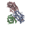


 Mass: 194.226 Da / Num. of mol.: 1 / Source method: obtained synthetically / Formula: C8H18O5 / Comment: precipitant*YM
 Mass: 106.120 Da / Num. of mol.: 6 / Source method: obtained synthetically / Formula: C4H10O3
 Mass: 65.409 Da / Num. of mol.: 4 / Source method: obtained synthetically / Formula: Zn
 Mass: 62.068 Da / Num. of mol.: 1 / Source method: obtained synthetically / Formula: C2H6O2
 Mass: 150.173 Da / Num. of mol.: 1 / Source method: obtained synthetically / Formula: C6H14O4
 Mass: 92.094 Da / Num. of mol.: 3 / Source method: obtained synthetically / Formula: C3H8O3
 Sample preparation
 / Beamline: BL11-1 / Wavelength: 0.91837, 0.97926
 Processing