 Movie
Movie Controller

[English] 日本語
 Yorodumi
- PDB-6sf4: Apo form of the ribonucleotide reductase NrdB protein from Leeuwe... -
+ Open data

- Basic information
| Entry | Database: PDB / ID: 6sf4 | ||||||||||||
|---|---|---|---|---|---|---|---|---|---|---|---|---|---|
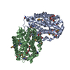
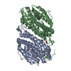
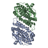
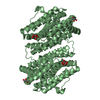
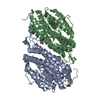
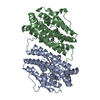
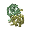
| Title | Apo form of the ribonucleotide reductase NrdB protein from Leeuwenhoekiella blandensis | ||||||||||||
 Components Components | Ribonucleoside-diphosphate reductase, beta subunit 1 | ||||||||||||
 Keywords Keywords | METAL BINDING PROTEIN / ribonucleotide reductase apoprotein manganese binding redox protein deoxyribonucleotide synthesis | ||||||||||||
| Function / homology |  Function and homology information Function and homology informationribonucleoside-diphosphate reductase / ribonucleoside-diphosphate reductase activity, thioredoxin disulfide as acceptor / deoxyribonucleotide biosynthetic process / ATP binding / metal ion binding Similarity search - Function | ||||||||||||
| Biological species |  Leeuwenhoekiella blandensis (bacteria) Leeuwenhoekiella blandensis (bacteria) | ||||||||||||
| Method |  X-RAY DIFFRACTION / SYNCHROTRON / MOLECULAR REPLACEMENT / Resolution: 1.7 Å X-RAY DIFFRACTION / SYNCHROTRON / MOLECULAR REPLACEMENT / Resolution: 1.7 Å | ||||||||||||
 Authors Authors | Hasan, M. / Rozman Grinberg, I. / Sjoberg, B.M. / Logan, D.T. | ||||||||||||
| Funding support |  Sweden, 3items Sweden, 3items
| ||||||||||||
 Citation Citation | Journal: J.Biol.Inorg.Chem. / Year: 2019 Title: Class Id ribonucleotide reductase utilizes a Mn2(IV,III) cofactor and undergoes large conformational changes on metal loading. Authors: Rozman Grinberg, I. / Berglund, S. / Hasan, M. / Lundin, D. / Ho, F.M. / Magnuson, A. / Logan, D.T. / Sjoberg, B.M. / Berggren, G. | ||||||||||||
| History |
|
- Structure visualization
| Structure viewer | Molecule: MolmilJmol/JSmol |
|---|
- Downloads & links
-Download
| PDBx/mmCIF format | 6sf4.cif.gz | 144.3 KB | Display | PDBx/mmCIF format |
|---|---|---|---|---|
| PDB format | pdb6sf4.ent.gz | 111.7 KB | Display | PDB format |
| PDBx/mmJSON format | 6sf4.json.gz | Tree view | PDBx/mmJSON format | |
| Others |  Other downloads Other downloads |
-Validation report
| Arichive directory | https://data.pdbj.org/pub/pdb/validation_reports/sf/6sf4ftp://data.pdbj.org/pub/pdb/validation_reports/sf/6sf4 | HTTPS FTP |
|---|
-Related structure data
| Related structure data |  6sf5C  1syyS S: Starting model for refinement C: citing same article ( |
|---|---|
| Similar structure data |


-Links
 PDBj
PDBj
- Assembly
| Deposited unit | 
| ||||||||
|---|---|---|---|---|---|---|---|---|---|
| 1 |
| ||||||||
| Unit cell |
|
-Components
| #1: Protein | Mass: 40577.906 Da / Num. of mol.: 2 Source method: isolated from a genetically manipulated source Source: (gene. exp.) Leeuwenhoekiella blandensis (strain CECT 7118 / CCUG 51940 / MED217) (bacteria)Strain: CECT 7118 / CCUG 51940 / MED217 / Gene: MED217_17135 / Production host: References: UniProt: A3XHF9, ribonucleoside-diphosphate reductase #2: Water | ChemComp-HOH / |  Mass: 18.015 Da / Num. of mol.: 486 / Source method: isolated from a natural source / Formula: H2O Mass: 18.015 Da / Num. of mol.: 486 / Source method: isolated from a natural source / Formula: H2O |
|---|
-Experimental details
-Experiment
| Experiment | Method: X-RAY DIFFRACTION / Number of used crystals: 1 |
|---|
- Sample preparation
| Crystal | Density Matthews: 2.47 Å3/Da / Density % sol: 50.18 % |
|---|---|
| Crystal grow | Temperature: 293 K / Method: vapor diffusion, sitting drop / pH: 7 Details: Protein at 7.5 mg/ml in buffer containing 50 mM Tris-HCl pH 7.8, 300 mM NaCl, 10% glycerol, 20 mM MgCl2 and 2 mM tris(2-carboxyethyl)phosphine (TCEP). Precipitant 2.4 M sodium malonate ...Details: Protein at 7.5 mg/ml in buffer containing 50 mM Tris-HCl pH 7.8, 300 mM NaCl, 10% glycerol, 20 mM MgCl2 and 2 mM tris(2-carboxyethyl)phosphine (TCEP). Precipitant 2.4 M sodium malonate dibasic monohydrate pH 7.0. Drops 200+200 nl. |
-Data collection
| Diffraction | Mean temperature: 100 K / Serial crystal experiment: N |
|---|---|
| Diffraction source | Source: SYNCHROTRON / Site: PETRA III, EMBL c/o DESY  / Beamline: P13 (MX1) / Wavelength: 0.85 Å / Beamline: P13 (MX1) / Wavelength: 0.85 Å |
| Detector | Type: DECTRIS PILATUS 6M / Detector: PIXEL / Date: Jul 14, 2016 |
| Radiation | Protocol: SINGLE WAVELENGTH / Monochromatic (M) / Laue (L): M / Scattering type: x-ray |
| Radiation wavelength | Wavelength: 0.85 Å / Relative weight: 1 |
| Reflection | Resolution: 1.7→49.94 Å / Num. obs: 74093 / % possible obs: 97.5 % / Redundancy: 17.5 % / Biso Wilson estimate: 32.5 Å2 / CC1/2: 0.999 / Rmerge(I) obs: 0.106 / Rpim(I) all: 0.035 / Net I/σ(I): 14.5 |
| Reflection shell | Resolution: 1.7→1.73 Å / Redundancy: 14.8 % / Rmerge(I) obs: 2.299 / Mean I/σ(I) obs: 1 / Num. unique obs: 3025 / CC1/2: 0.538 / Rpim(I) all: 0.854 / % possible all: 76.2 |
- Processing
| Software |
| ||||||||||||||||||||
|---|---|---|---|---|---|---|---|---|---|---|---|---|---|---|---|---|---|---|---|---|---|
| Refinement | Method to determine structure: MOLECULAR REPLACEMENT Starting model: 1SYY Resolution: 1.7→49.9 Å / Cor.coef. Fo:Fc: 0.96 / Cor.coef. Fo:Fc free: 0.954 / SU R Cruickshank DPI: 0.096 / Cross valid method: THROUGHOUT / SU R Blow DPI: 0.1 / SU Rfree Blow DPI: 0.091 / SU Rfree Cruickshank DPI: 0.089
| ||||||||||||||||||||
| Displacement parameters | Biso mean: 35.15 Å2
| ||||||||||||||||||||
| Refine analyze | Luzzati coordinate error obs: 0.22 Å | ||||||||||||||||||||
| Refinement step | Cycle: LAST / Resolution: 1.7→49.9 Å
|
