[English] 日本語
Yorodumi
- PDB-6sf5: Mn-containing form of the ribonucleotide reductase NrdB protein f... -

+
Open data


ID or keywords:

Loading...

-
Basic information

Entry
Database: PDB / ID: 6sf5
TitleMn-containing form of the ribonucleotide reductase NrdB protein from Leeuwenhoekiella blandensis
ComponentsRibonucleoside-diphosphate reductase, beta subunit 1
Keywords
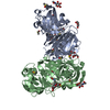
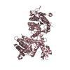
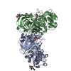
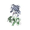
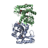
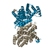
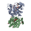
METAL BINDING PROTEIN / ribonucleotide reductase apoprotein manganese binding redox protein deoxyribonucleotide synthesis
Function / homology
Function and homology information


ribonucleoside-diphosphate reductase / ribonucleoside-diphosphate reductase activity, thioredoxin disulfide as acceptor / deoxyribonucleotide biosynthetic process / ATP binding / metal ion binding
Similarity search - Function
ATP-cone domain / ATP cone domain / ATP-cone domain profile. / Ribonucleotide reductase small subunit / Ribonucleotide reductase small subunit family / Ribonucleotide reductase, small chain / Ribonucleotide reductase-like / Ferritin-like superfamily
Similarity search - Domain/homology
: / ribonucleoside-diphosphate reductase
Similarity search - Component
Biological speciesLeeuwenhoekiella blandensis (bacteria)
MethodX-RAY DIFFRACTION / SYNCHROTRON / MOLECULAR REPLACEMENT / Resolution: 1.9 Å
AuthorsHasan, M. / Rozman Grinberg, I. / Sjoberg, B.M. / Logan, D.T.
Funding support Sweden, 3items
OrganizationGrant numberCountry
Swedish Research Council2016-04855 Sweden
Swedish Research Council2016-01920 Sweden
Wenner-Gren Foundation Sweden
CitationJournal: J.Biol.Inorg.Chem. / Year: 2019
Title: Class Id ribonucleotide reductase utilizes a Mn2(IV,III) cofactor and undergoes large conformational changes on metal loading.
Authors: Rozman Grinberg, I. / Berglund, S. / Hasan, M. / Lundin, D. / Ho, F.M. / Magnuson, A. / Logan, D.T. / Sjoberg, B.M. / Berggren, G.
History
DepositionAug 1, 2019Deposition site: PDBE / Processing site: PDBE
Revision 1.0Aug 28, 2019Provider: repository / Type: Initial release
Revision 1.1Oct 2, 2019Group: Data collection / Database references / Category: citation
Item: _citation.journal_volume / _citation.page_first / _citation.page_last
Revision 1.2Nov 20, 2019Group: Derived calculations / Category: pdbx_struct_conn_angle / struct_conn
Revision 1.3Jan 24, 2024Group: Data collection / Database references ...Data collection / Database references / Derived calculations / Refinement description
Category: chem_comp_atom / chem_comp_bond ...chem_comp_atom / chem_comp_bond / database_2 / pdbx_initial_refinement_model / struct_conn
Item: _database_2.pdbx_DOI / _database_2.pdbx_database_accession ..._database_2.pdbx_DOI / _database_2.pdbx_database_accession / _struct_conn.pdbx_dist_value / _struct_conn.ptnr1_auth_asym_id / _struct_conn.ptnr1_auth_comp_id / _struct_conn.ptnr1_auth_seq_id / _struct_conn.ptnr1_label_asym_id / _struct_conn.ptnr1_label_atom_id / _struct_conn.ptnr1_label_comp_id / _struct_conn.ptnr1_label_seq_id / _struct_conn.ptnr2_auth_asym_id / _struct_conn.ptnr2_auth_comp_id / _struct_conn.ptnr2_auth_seq_id / _struct_conn.ptnr2_label_asym_id / _struct_conn.ptnr2_label_atom_id / _struct_conn.ptnr2_label_comp_id

-
Structure visualization

Structure viewerMolecule:
MolmilJmol/JSmol

Downloads & links

-
Assembly

Deposited unit
A: Ribonucleoside-diphosphate reductase, beta subunit 1
B: Ribonucleoside-diphosphate reductase, beta subunit 1
hetero molecules


Theoretical massNumber of molelcules
Total (without water)81,3766
Polymers81,1562
Non-polymers2204
Water6,972387
1


  • Idetical with deposited unit
  • defined by author&software
  • Evidence: gel filtration
TypeNameSymmetry operationNumber
identity operation1_555x,y,z1
Buried area2600 Å2
ΔGint-36 kcal/mol
Surface area24940 Å2
MethodPISA
Unit cell
Length a, b, c (Å)57.06, 80.602, 158.228
Angle α, β, γ (deg.)90, 90, 90
Int Tables number19
Space group name H-MP212121

-
Components

#1: Protein Ribonucleoside-diphosphate reductase, beta subunit 1


Mass: 40577.906 Da / Num. of mol.: 2
Source method: isolated from a genetically manipulated source
Source: (gene. exp.) Leeuwenhoekiella blandensis (bacteria) / Gene: MED217_17135 / Production host: Escherichia coli BL21(DE3) (bacteria) / Variant (production host): /pET28a(+)
References: UniProt: A3XHF9, ribonucleoside-diphosphate reductase
#2: Chemical
ChemComp-MN / MANGANESE (II) ION


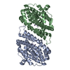
Mass: 54.938 Da / Num. of mol.: 4 / Source method: obtained synthetically / Formula: Mn / Feature type: SUBJECT OF INVESTIGATION
#3: Water ChemComp-HOH / water


Mass: 18.015 Da / Num. of mol.: 387 / Source method: isolated from a natural source / Formula: H2O
Has ligand of interestY

-
Experimental details

-
Experiment

ExperimentMethod: X-RAY DIFFRACTION / Number of used crystals: 1

-
Sample preparation

CrystalDensity Matthews: 2.24 Å3/Da / Density % sol: 45.13 %
Crystal growTemperature: 293 K / Method: vapor diffusion, sitting drop / pH: 5.5
Details: Protein at 7.5 mg/ml in 50 mM Tris-HCl pH 7.8, 300 mM NaCl, 10% glycerol, 20 mM MgCl2, 2 mM tris(2-carboxyethyl)phosphine (TCEP) and 10 mM MnCl2. 200+200 nl sitting drops. Precipitant 0.1 M ...Details: Protein at 7.5 mg/ml in 50 mM Tris-HCl pH 7.8, 300 mM NaCl, 10% glycerol, 20 mM MgCl2, 2 mM tris(2-carboxyethyl)phosphine (TCEP) and 10 mM MnCl2. 200+200 nl sitting drops. Precipitant 0.1 M Bis-Tris pH 5.5, 25% w/v polyethylene glycol 3350.

-
Data collection

DiffractionMean temperature: 100 K / Serial crystal experiment: N
Diffraction sourceSource: SYNCHROTRON / Site: PETRA III, EMBL c/o DESY / Beamline: P14 (MX2) / Wavelength: 0.9763 Å
DetectorType: DECTRIS EIGER X 16M / Detector: PIXEL / Date: Nov 24, 2017
RadiationProtocol: SINGLE WAVELENGTH / Monochromatic (M) / Laue (L): M / Scattering type: x-ray
Radiation wavelengthWavelength: 0.9763 Å / Relative weight: 1
ReflectionResolution: 1.9→80.6 Å / Num. obs: 17348 / % possible obs: 99.9 % / Redundancy: 6.5 % / CC1/2: 0.999 / Rmerge(I) obs: 0.062 / Rpim(I) all: 0.028 / Net I/σ(I): 13.5
Reflection shellResolution: 1.9→1.94 Å / Rmerge(I) obs: 1.137 / Mean I/σ(I) obs: 1.2 / Num. unique obs: 3670 / CC1/2: 0.475 / Rpim(I) all: 0.658 / % possible all: 99.5

-
Processing

Software
NameVersionClassification
BUSTER2.11.7refinement
XDSdata reduction
Aimlessdata scaling
PHASERphasing
RefinementMethod to determine structure: MOLECULAR REPLACEMENT
Starting model: 6SF4
Resolution: 1.9→71.82 Å / Cor.coef. Fo:Fc: 0.957 / Cor.coef. Fo:Fc free: 0.957 / SU R Cruickshank DPI: 0.126 / Cross valid method: THROUGHOUT / SU R Blow DPI: 0.13 / SU Rfree Blow DPI: 0.113 / SU Rfree Cruickshank DPI: 0.112
RfactorNum. reflection% reflectionSelection details
Rfree0.195 2895 -RANDOM
Rwork0.175 ---
obs0.176 58284 99.9 %-
Displacement parametersBiso mean: 51.11 Å2
Baniso -1Baniso -2Baniso -3
1--13.5696 Å20 Å20 Å2
2--7.562 Å20 Å2
3---6.0076 Å2
Refine analyzeLuzzati coordinate error obs: 0.23 Å
Refinement stepCycle: LAST / Resolution: 1.9→71.82 Å
ProteinNucleic acidLigandSolventTotal
Num. atoms4893 0 4 387 5284
Refine LS restraints
Refine-IDTypeDev idealNumberRestraint functionWeight
X-RAY DIFFRACTIONt_bond_d0.015042HARMONIC2
X-RAY DIFFRACTIONt_angle_deg0.946818HARMONIC2
X-RAY DIFFRACTIONt_dihedral_angle_d1816SINUSOIDAL2
X-RAY DIFFRACTIONt_incorr_chiral_ct
X-RAY DIFFRACTIONt_pseud_angle
X-RAY DIFFRACTIONt_trig_c_planes
X-RAY DIFFRACTIONt_gen_planes866HARMONIC5
X-RAY DIFFRACTIONt_it5042HARMONIC20
X-RAY DIFFRACTIONt_nbd1SEMIHARMONIC5
X-RAY DIFFRACTIONt_omega_torsion2.82
X-RAY DIFFRACTIONt_other_torsion17.95
X-RAY DIFFRACTIONt_improper_torsion
X-RAY DIFFRACTIONt_chiral_improper_torsion659SEMIHARMONIC5
X-RAY DIFFRACTIONt_sum_occupancies
X-RAY DIFFRACTIONt_utility_distance
X-RAY DIFFRACTIONt_utility_angle
X-RAY DIFFRACTIONt_utility_torsion
X-RAY DIFFRACTIONt_ideal_dist_contact6364SEMIHARMONIC4
LS refinement shellResolution: 1.9→1.91 Å
RfactorNum. reflection% reflection
Rfree0.3088 51 -
Rwork0.2221 1115 -
all0.2257 1166 -
obs--98.37 %
Refinement TLS params.

Refine-ID: X-RAY DIFFRACTION

IDL112)L122)L132)L222)L232)L332)S11 (Å °)S12 (Å °)S13 (Å °)S21 (Å °)S22 (Å °)S23 (Å °)S31 (Å °)S32 (Å °)S33 (Å °)T112)T122)T132)T222)T232)T332)Origin x (Å)Origin y (Å)Origin z (Å)
11.0701-0.71460.50722.04310.151.79060.0246-0.1438-0.116-0.14380.00730.0343-0.1160.0343-0.0319-0.1329-0.02880.0345-0.1074-0.0085-0.078827.3507-1.922155.943
21.41170.88490.59232.07360.8542.30580.17140.52490.07160.5249-0.176-0.42390.0716-0.42390.0046-0.00080.02440.0742-0.08720.0118-0.195920.09918.4655189.872
Refinement TLS group
IDRefine-IDRefine TLS-IDSelection details
1X-RAY DIFFRACTION1{ A|* }
2X-RAY DIFFRACTION2{ B|* }

+
About Yorodumi

-
News

-
Feb 9, 2022. New format data for meta-information of EMDB entries

New format data for meta-information of EMDB entries

  • Version 3 of the EMDB header file is now the official format.
  • The previous official version 1.9 will be removed from the archive.

Related info.:EMDB header

External links:wwPDB to switch to version 3 of the EMDB data model

-
Aug 12, 2020. Covid-19 info

Covid-19 info

URL: https://pdbjlvh1.pdbj.org/emnavi/covid19.php

New page: Covid-19 featured information page in EM Navigator.

Related info.:Covid-19 info / Mar 5, 2020. Novel coronavirus structure data

+
Mar 5, 2020. Novel coronavirus structure data

Novel coronavirus structure data

Related info.:Yorodumi Speices / Aug 12, 2020. Covid-19 info

External links:COVID-19 featured content - PDBj / Molecule of the Month (242):Coronavirus Proteases

+
Jan 31, 2019. EMDB accession codes are about to change! (news from PDBe EMDB page)

EMDB accession codes are about to change! (news from PDBe EMDB page)

  • The allocation of 4 digits for EMDB accession codes will soon come to an end. Whilst these codes will remain in use, new EMDB accession codes will include an additional digit and will expand incrementally as the available range of codes is exhausted. The current 4-digit format prefixed with “EMD-” (i.e. EMD-XXXX) will advance to a 5-digit format (i.e. EMD-XXXXX), and so on. It is currently estimated that the 4-digit codes will be depleted around Spring 2019, at which point the 5-digit format will come into force.
  • The EM Navigator/Yorodumi systems omit the EMD- prefix.

Related info.:Q: What is EMD? / ID/Accession-code notation in Yorodumi/EM Navigator

External links:EMDB Accession Codes are Changing Soon! / Contact to PDBj

+
Jul 12, 2017. Major update of PDB

Major update of PDB

  • wwPDB released updated PDB data conforming to the new PDBx/mmCIF dictionary.
  • This is a major update changing the version number from 4 to 5, and with Remediation, in which all the entries are updated.
  • In this update, many items about electron microscopy experimental information are reorganized (e.g. em_software).
  • Now, EM Navigator and Yorodumi are based on the updated data.

External links:wwPDB Remediation / Enriched Model Files Conforming to OneDep Data Standards Now Available in the PDB FTP Archive

-
Yorodumi

Thousand views of thousand structures

  • Yorodumi is a browser for structure data from EMDB, PDB, SASBDB, etc.
  • This page is also the successor to EM Navigator detail page, and also detail information page/front-end page for Omokage search.
  • The word "yorodu" (or yorozu) is an old Japanese word meaning "ten thousand". "mi" (miru) is to see.

Related info.:EMDB / PDB / SASBDB / Comparison of 3 databanks / Yorodumi Search / Aug 31, 2016. New EM Navigator & Yorodumi / Yorodumi Papers / Jmol/JSmol / Function and homology information / Changes in new EM Navigator and Yorodumi

Read more