 Movie
Movie Controller

[English] 日本語
 Yorodumi
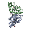
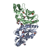
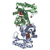
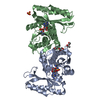
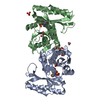
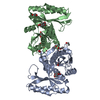
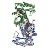
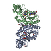
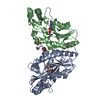
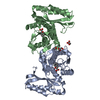
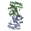
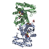
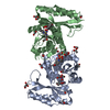
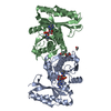
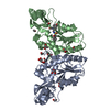
- PDB-4ddk: Pantothenate synthetase in complex with 1,3-benzodioxole-5-carbox... -
+ Open data

- Basic information
| Entry | Database: PDB / ID: 4ddk | ||||||
|---|---|---|---|---|---|---|---|
| Title | Pantothenate synthetase in complex with 1,3-benzodioxole-5-carboxylic acid | ||||||
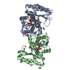
 Components Components | Pantothenate synthetase | ||||||
 Keywords Keywords | ligase/ligase inhibitor / Fragment-based drug discovery / Alpha Beta/Adenine nucleotide alpha hydrolase-like / pantoate and b-Ala binding / ligase-ligase inhibitor complex | ||||||
| Function / homology |  Function and homology information Function and homology information: / pantoate-beta-alanine ligase (AMP-forming) / pantoate-beta-alanine ligase activity / pantothenate biosynthetic process / manganese ion binding / magnesium ion binding / ATP binding / metal ion binding / cytoplasm / cytosol Similarity search - Function | ||||||
| Biological species |   Mycobacterium tuberculosis (bacteria) Mycobacterium tuberculosis (bacteria) | ||||||
| Method |  X-RAY DIFFRACTION / SYNCHROTRON / MOLECULAR REPLACEMENT / Resolution: 1.75 Å X-RAY DIFFRACTION / SYNCHROTRON / MOLECULAR REPLACEMENT / Resolution: 1.75 Å | ||||||
 Authors Authors | Silvestre, H.L. | ||||||
 Citation Citation | Journal: Proc.Natl.Acad.Sci.USA / Year: 2013 Title: Integrated biophysical approach to fragment screening and validation for fragment-based lead discovery. Authors: Silvestre, H.L. / Blundell, T.L. / Abell, C. / Ciulli, A. | ||||||
| History |
|
- Structure visualization
| Structure viewer | Molecule: MolmilJmol/JSmol |
|---|
- Downloads & links
-Download
| PDBx/mmCIF format | 4ddk.cif.gz | 133.4 KB | Display | PDBx/mmCIF format |
|---|---|---|---|---|
| PDB format | pdb4ddk.ent.gz | 102.9 KB | Display | PDB format |
| PDBx/mmJSON format | 4ddk.json.gz | Tree view | PDBx/mmJSON format | |
| Others |  Other downloads Other downloads |
-Validation report
| Arichive directory | https://data.pdbj.org/pub/pdb/validation_reports/dd/4ddkftp://data.pdbj.org/pub/pdb/validation_reports/dd/4ddk | HTTPS FTP |
|---|
-Related structure data
| Related structure data |  4ddhC  4ddmC  4de5C 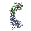 4ef6C  4efkC  4fzjC  4g5fC  4g5yC  3covS C: citing same article ( S: Starting model for refinement |
|---|---|
| Similar structure data |


-Links
 PDBj
PDBj


- Assembly
| Deposited unit | 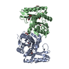
| ||||||||
|---|---|---|---|---|---|---|---|---|---|
| 1 |
| ||||||||
| 2 | 
| ||||||||
| Unit cell |
|
-Components
-Protein , 1 types, 2 molecules AB
| #1: Protein | Mass: 31571.178 Da / Num. of mol.: 2 / Mutation: T2A, E77G Source method: isolated from a genetically manipulated source Source: (gene. exp.) Mycobacterium tuberculosis (bacteria) / Strain: H37Rv / Gene: MT3707, MTCY07H7B.20, panC, Rv3602c / Plasmid: pET30b / Production host: References: UniProt: P0A5R0, UniProt: P9WIL5*PLUS, pantoate-beta-alanine ligase (AMP-forming) |
|---|
-Non-polymers , 5 types, 575 molecules 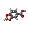


| #2: Chemical |  Mass: 166.131 Da / Num. of mol.: 3 / Source method: obtained synthetically / Formula: C8H6O4 Mass: 166.131 Da / Num. of mol.: 3 / Source method: obtained synthetically / Formula: C8H6O4#3: Chemical | ChemComp-GOL /  Mass: 92.094 Da / Num. of mol.: 4 / Source method: obtained synthetically / Formula: C3H8O3 Mass: 92.094 Da / Num. of mol.: 4 / Source method: obtained synthetically / Formula: C3H8O3#4: Chemical |  Mass: 46.068 Da / Num. of mol.: 2 / Source method: obtained synthetically / Formula: C2H6O Mass: 46.068 Da / Num. of mol.: 2 / Source method: obtained synthetically / Formula: C2H6O#5: Chemical | ChemComp-EDO / |  Mass: 62.068 Da / Num. of mol.: 1 / Source method: obtained synthetically / Formula: C2H6O2 Mass: 62.068 Da / Num. of mol.: 1 / Source method: obtained synthetically / Formula: C2H6O2#6: Water | ChemComp-HOH / | Mass: 18.015 Da / Num. of mol.: 565 / Source method: isolated from a natural source / Formula: H2O |
|---|
-Experimental details
-Experiment
| Experiment | Method: X-RAY DIFFRACTION / Number of used crystals: 1 |
|---|
- Sample preparation
| Crystal | Density Matthews: 2.11 Å3/Da / Density % sol: 41.62 % |
|---|---|
| Crystal grow | Temperature: 294 K / Method: vapor diffusion, hanging drop / pH: 8 Details: 12% PEG 3000, 150 mM Li2SO4, 0.1 M imidazole, 2% ethanol, 10% ethylene glycol, 20 mM MgCl2, pH 8.0, VAPOR DIFFUSION, HANGING DROP, temperature 294K |
-Data collection
| Diffraction | Mean temperature: 100 K |
|---|---|
| Diffraction source | Source: SYNCHROTRON / Site: ESRF  / Beamline: ID14-1 / Wavelength: 0.93 Å / Beamline: ID14-1 / Wavelength: 0.93 Å |
| Detector | Type: ADSC QUANTUM 210 / Detector: CCD / Date: Feb 16, 2008 |
| Radiation | Monochromator: Diamond (111), Ge(220) Sagitally focusing Ge(220) Protocol: SINGLE WAVELENGTH / Monochromatic (M) / Laue (L): M / Scattering type: x-ray |
| Radiation wavelength | Wavelength: 0.93 Å / Relative weight: 1 |
| Reflection | Resolution: 1.75→26.854 Å / Num. all: 142967 / Num. obs: 49952 / % possible obs: 91.5 % / Redundancy: 2.9 % / Biso Wilson estimate: 16.39 Å2 / Rmerge(I) obs: 0.077 / Rsym value: 0.077 / Net I/σ(I): 11.6 |
| Reflection shell | Resolution: 1.75→1.84 Å / Redundancy: 2.8 % / Rmerge(I) obs: 0.422 / Mean I/σ(I) obs: 2.8 / Num. unique all: 7129 / % possible all: 89.6 |
- Processing
| Software |
| ||||||||||||||||||||||||||||||||||||||||||||||||||||||||||||||||||||||||||||||||||||||||||||||||||||||||||||||||||||||||||||||||||||||||||||||||||||||||||||||||||||||||||
|---|---|---|---|---|---|---|---|---|---|---|---|---|---|---|---|---|---|---|---|---|---|---|---|---|---|---|---|---|---|---|---|---|---|---|---|---|---|---|---|---|---|---|---|---|---|---|---|---|---|---|---|---|---|---|---|---|---|---|---|---|---|---|---|---|---|---|---|---|---|---|---|---|---|---|---|---|---|---|---|---|---|---|---|---|---|---|---|---|---|---|---|---|---|---|---|---|---|---|---|---|---|---|---|---|---|---|---|---|---|---|---|---|---|---|---|---|---|---|---|---|---|---|---|---|---|---|---|---|---|---|---|---|---|---|---|---|---|---|---|---|---|---|---|---|---|---|---|---|---|---|---|---|---|---|---|---|---|---|---|---|---|---|---|---|---|---|---|---|---|---|---|
| Refinement | Method to determine structure: MOLECULAR REPLACEMENT Starting model: pdb entry 3COV Resolution: 1.75→26.854 Å / Cor.coef. Fo:Fc: 0.951 / Cor.coef. Fo:Fc free: 0.914 / SU B: 3.617 / SU ML: 0.113 / Cross valid method: THROUGHOUT / ESU R: 0.143 / ESU R Free: 0.143 / Stereochemistry target values: MAXIMUM LIKELIHOOD / Details: HYDROGENS HAVE BEEN ADDED IN THE RIDING POSITIONS
| ||||||||||||||||||||||||||||||||||||||||||||||||||||||||||||||||||||||||||||||||||||||||||||||||||||||||||||||||||||||||||||||||||||||||||||||||||||||||||||||||||||||||||
| Solvent computation | Ion probe radii: 0.8 Å / Shrinkage radii: 0.8 Å / VDW probe radii: 1.2 Å / Solvent model: MASK | ||||||||||||||||||||||||||||||||||||||||||||||||||||||||||||||||||||||||||||||||||||||||||||||||||||||||||||||||||||||||||||||||||||||||||||||||||||||||||||||||||||||||||
| Displacement parameters | Biso mean: 20.653 Å2
| ||||||||||||||||||||||||||||||||||||||||||||||||||||||||||||||||||||||||||||||||||||||||||||||||||||||||||||||||||||||||||||||||||||||||||||||||||||||||||||||||||||||||||
| Refinement step | Cycle: LAST / Resolution: 1.75→26.854 Å
| ||||||||||||||||||||||||||||||||||||||||||||||||||||||||||||||||||||||||||||||||||||||||||||||||||||||||||||||||||||||||||||||||||||||||||||||||||||||||||||||||||||||||||
| Refine LS restraints |
| ||||||||||||||||||||||||||||||||||||||||||||||||||||||||||||||||||||||||||||||||||||||||||||||||||||||||||||||||||||||||||||||||||||||||||||||||||||||||||||||||||||||||||
| LS refinement shell | Resolution: 1.75→1.795 Å / Total num. of bins used: 20
|
