 Movie
Movie Controller

[English] 日本語
 Yorodumi
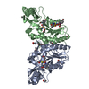
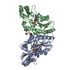
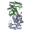
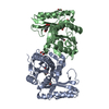
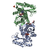
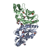
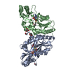
- PDB-1n2i: Crystal Structure of Pantothenate Synthetase from M. tuberculosis... -
+ Open data

- Basic information
| Entry | Database: PDB / ID: 1n2i | ||||||
|---|---|---|---|---|---|---|---|
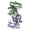
| Title | Crystal Structure of Pantothenate Synthetase from M. tuberculosis in complex with a reaction intermediate, pantoyl adenylate, different occupancies of pantoyl adenylate | ||||||
 Components Components | pantothenate synthetase | ||||||
 Keywords Keywords | LIGASE / Structural genomics / Rossmann fold / dimer / intersubunit beta sheet / PSI / Protein Structure Initiative / TB Structural Genomics Consortium / TBSGC | ||||||
| Function / homology |  Function and homology information Function and homology information: / pantoate-beta-alanine ligase (AMP-forming) / pantoate-beta-alanine ligase activity / pantothenate biosynthetic process / manganese ion binding / magnesium ion binding / ATP binding / metal ion binding / cytosol / cytoplasm Similarity search - Function | ||||||
| Biological species |   Mycobacterium tuberculosis (bacteria) Mycobacterium tuberculosis (bacteria) | ||||||
| Method |  X-RAY DIFFRACTION / rigid body refinement into new data / Resolution: 1.7 Å X-RAY DIFFRACTION / rigid body refinement into new data / Resolution: 1.7 Å | ||||||
 Authors Authors | Wang, S. / Eisenberg, D. / TB Structural Genomics Consortium (TBSGC) | ||||||
 Citation Citation | Journal: Protein Sci. / Year: 2003 Title: Crystal structures of a pantothenate synthetase from M. tuberculosis and its complexes with substrates and a reaction intermediate Authors: Wang, S. / Eisenberg, D. | ||||||
| History |
|
- Structure visualization
| Structure viewer | Molecule: MolmilJmol/JSmol |
|---|
- Downloads & links
-Download
| PDBx/mmCIF format | 1n2i.cif.gz | 127.9 KB | Display | PDBx/mmCIF format |
|---|---|---|---|---|
| PDB format | pdb1n2i.ent.gz | 99.7 KB | Display | PDB format |
| PDBx/mmJSON format | 1n2i.json.gz | Tree view | PDBx/mmJSON format | |
| Others |  Other downloads Other downloads |
-Validation report
| Arichive directory | https://data.pdbj.org/pub/pdb/validation_reports/n2/1n2iftp://data.pdbj.org/pub/pdb/validation_reports/n2/1n2i | HTTPS FTP |
|---|
-Related structure data
| Related structure data |  1mopSC  1n2bC  1n2eC  1n2gC  1n2hC 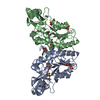 1n2jC  1n2oC S: Starting model for refinement C: citing same article ( |
|---|---|
| Similar structure data | |
| Other databases |


-Links
 PDBj
PDBj


- Assembly
| Deposited unit | 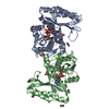
| ||||||||
|---|---|---|---|---|---|---|---|---|---|
| 1 |
| ||||||||
| Unit cell |
| ||||||||
| Details | The biological assembly is a dimer |
-Components
-Protein , 1 types, 2 molecules AB
| #1: Protein | Mass: 31500.100 Da / Num. of mol.: 2 / Mutation: T2A, E77G Source method: isolated from a genetically manipulated source Source: (gene. exp.) Mycobacterium tuberculosis (bacteria) / Gene: panC / Plasmid: pET30a / Species (production host): Escherichia coli / Production host: References: UniProt: P0A5R0, UniProt: P9WIL5*PLUS, pantoate-beta-alanine ligase (AMP-forming) |
|---|
-Non-polymers , 5 types, 412 molecules 
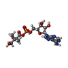


| #2: Chemical |  Mass: 96.063 Da / Num. of mol.: 3 / Source method: obtained synthetically / Formula: SO4 Mass: 96.063 Da / Num. of mol.: 3 / Source method: obtained synthetically / Formula: SO4#3: Chemical |  Mass: 477.363 Da / Num. of mol.: 2 / Source method: obtained synthetically / Formula: C16H24N5O10P Mass: 477.363 Da / Num. of mol.: 2 / Source method: obtained synthetically / Formula: C16H24N5O10P#4: Chemical | ChemComp-GOL / |  Mass: 92.094 Da / Num. of mol.: 1 / Source method: obtained synthetically / Formula: C3H8O3 Mass: 92.094 Da / Num. of mol.: 1 / Source method: obtained synthetically / Formula: C3H8O3#5: Chemical | ChemComp-EOH /  Mass: 46.068 Da / Num. of mol.: 5 / Source method: obtained synthetically / Formula: C2H6O Mass: 46.068 Da / Num. of mol.: 5 / Source method: obtained synthetically / Formula: C2H6O#6: Water | ChemComp-HOH / | Mass: 18.015 Da / Num. of mol.: 401 / Source method: isolated from a natural source / Formula: H2O |
|---|
-Experimental details
-Experiment
| Experiment | Method: X-RAY DIFFRACTION / Number of used crystals: 1 |
|---|
- Sample preparation
| Crystal | Density Matthews: 2.18 Å3/Da / Density % sol: 43.62 % |
|---|---|
| Crystal grow | Temperature: 293 K / Method: vapor diffusion, hanging drop / pH: 8 Details: PEG3000, lithium sulfate, magnesium chloride, imidazol, ethanol, glycerol, pH 8.0, VAPOR DIFFUSION, HANGING DROP, temperature 293K |
-Data collection
| Diffraction | Mean temperature: 100 K |
|---|---|
| Diffraction source | Source: ROTATING ANODE / Type: RIGAKU FR-D / Wavelength: 1.5418 |
| Detector | Type: RIGAKU RAXIS IV++ / Detector: IMAGE PLATE / Date: Jun 3, 2002 / Details: mirrors |
| Radiation | Monochromator: Ni Filter / Protocol: SINGLE WAVELENGTH / Monochromatic (M) / Laue (L): M / Scattering type: x-ray |
| Radiation wavelength | Wavelength: 1.5418 Å / Relative weight: 1 |
| Reflection | Resolution: 1.7→50 Å / Num. all: 58966 / Num. obs: 58966 / % possible obs: 98.8 % / Observed criterion σ(I): -3 / Redundancy: 7.5 % / Biso Wilson estimate: 25.4 Å2 / Rmerge(I) obs: 0.077 / Rsym value: 0.077 / Net I/σ(I): 23.7 |
| Reflection shell | Resolution: 1.7→1.76 Å / Redundancy: 5.7 % / Rmerge(I) obs: 0.489 / Mean I/σ(I) obs: 2.9 / Num. unique all: 5586 / Rsym value: 0.489 / % possible all: 94.1 |
- Processing
| Software |
| ||||||||||||||||||||||||||||||||||||||||||||||||||||||||||||||||||||||||||||||||
|---|---|---|---|---|---|---|---|---|---|---|---|---|---|---|---|---|---|---|---|---|---|---|---|---|---|---|---|---|---|---|---|---|---|---|---|---|---|---|---|---|---|---|---|---|---|---|---|---|---|---|---|---|---|---|---|---|---|---|---|---|---|---|---|---|---|---|---|---|---|---|---|---|---|---|---|---|---|---|---|---|---|
| Refinement | Method to determine structure: rigid body refinement into new data Starting model: PDB entry 1MOP Resolution: 1.7→19.8 Å / Rfactor Rfree error: 0.003 / Isotropic thermal model: RESTRAINED / Cross valid method: THROUGHOUT / σ(F): 0 / Stereochemistry target values: Engh & Huber Details: The occupancies for the ligands were not refined. They are inferred from the temperature factors of the ligand atoms, which were refined with an occupancy of 1.00 for all atoms. The pantoyl ...Details: The occupancies for the ligands were not refined. They are inferred from the temperature factors of the ligand atoms, which were refined with an occupancy of 1.00 for all atoms. The pantoyl adenylate molecule in subunit B has an average temperature factor similar to that of the nearby protein atoms in the active site. However, that in subunit A has a much higher average temperature factor.
| ||||||||||||||||||||||||||||||||||||||||||||||||||||||||||||||||||||||||||||||||
| Solvent computation | Solvent model: FLAT MODEL / Bsol: 48.7665 Å2 / ksol: 0.373287 e/Å3 | ||||||||||||||||||||||||||||||||||||||||||||||||||||||||||||||||||||||||||||||||
| Displacement parameters | Biso mean: 26.6 Å2
| ||||||||||||||||||||||||||||||||||||||||||||||||||||||||||||||||||||||||||||||||
| Refine analyze |
| ||||||||||||||||||||||||||||||||||||||||||||||||||||||||||||||||||||||||||||||||
| Refinement step | Cycle: LAST / Resolution: 1.7→19.8 Å
| ||||||||||||||||||||||||||||||||||||||||||||||||||||||||||||||||||||||||||||||||
| Refine LS restraints |
| ||||||||||||||||||||||||||||||||||||||||||||||||||||||||||||||||||||||||||||||||
| LS refinement shell | Resolution: 1.7→1.81 Å / Rfactor Rfree error: 0.012 / Total num. of bins used: 6
| ||||||||||||||||||||||||||||||||||||||||||||||||||||||||||||||||||||||||||||||||
| Xplor file |
|
