 Movie
Movie Controller

[English] 日本語
 Yorodumi
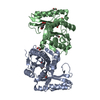
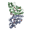
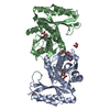
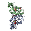
- PDB-1n2g: Crystal Structure of a Pantothenate Synthetase from M. tuberculos... -
+ Open data

- Basic information
| Entry | Database: PDB / ID: 1n2g | ||||||
|---|---|---|---|---|---|---|---|
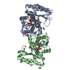
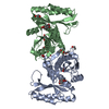
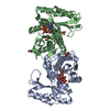
| Title | Crystal Structure of a Pantothenate Synthetase from M. tuberculosis in complex with AMPCPP | ||||||
 Components Components | Pantothenate synthetase | ||||||
 Keywords Keywords | LIGASE / Structural genomics / Rossmann fold / dimer / intersubunit beta sheet / PSI / Protein Structure Initiative / TB Structural Genomics Consortium / TBSGC | ||||||
| Function / homology |  Function and homology information Function and homology information: / pantoate-beta-alanine ligase (AMP-forming) / pantoate-beta-alanine ligase activity / pantothenate biosynthetic process / manganese ion binding / magnesium ion binding / ATP binding / metal ion binding / cytosol / cytoplasm Similarity search - Function | ||||||
| Biological species |   Mycobacterium tuberculosis (bacteria) Mycobacterium tuberculosis (bacteria) | ||||||
| Method |  X-RAY DIFFRACTION / rigid body refinement into new data / Resolution: 1.8 Å X-RAY DIFFRACTION / rigid body refinement into new data / Resolution: 1.8 Å | ||||||
 Authors Authors | Wang, S. / Eisenberg, D. / TB Structural Genomics Consortium (TBSGC) | ||||||
 Citation Citation | Journal: Protein Sci. / Year: 2003 Title: Crystal structures of a pantothenate synthetase from M. tuberculosis and its complexes with substrates and a reaction intermediate Authors: Wang, S. / Eisenberg, D. | ||||||
| History |
| ||||||
| Remark 999 | SEQUENCE RESIDUE 2 IS AN ALANINE IN THE SEQUENCE. MUTATION OF THIS RESIDUE WAS NECESSARY TO ... SEQUENCE RESIDUE 2 IS AN ALANINE IN THE SEQUENCE. MUTATION OF THIS RESIDUE WAS NECESSARY TO GENERATE AN NCOI RESTRICTION SITE FOR CLONING INTO THE EXPRESSION VECTOR PET30A. THE LAST 9 RESIDUES AFTER ARG300 WERE CLEAVED OFF BY ENTEROKINASE DIGESTION (UNINTENTIONALLY), WHICH WAS CARRIED OUT TO CLEAVE THE N-TERMINAL TAG FROM THE RECOMBINANT PROTEIN. |
- Structure visualization
| Structure viewer | Molecule: MolmilJmol/JSmol |
|---|
- Downloads & links
-Download
| PDBx/mmCIF format | 1n2g.cif.gz | 127.6 KB | Display | PDBx/mmCIF format |
|---|---|---|---|---|
| PDB format | pdb1n2g.ent.gz | 98.8 KB | Display | PDB format |
| PDBx/mmJSON format | 1n2g.json.gz | Tree view | PDBx/mmJSON format | |
| Others |  Other downloads Other downloads |
-Validation report
| Arichive directory | https://data.pdbj.org/pub/pdb/validation_reports/n2/1n2gftp://data.pdbj.org/pub/pdb/validation_reports/n2/1n2g | HTTPS FTP |
|---|
-Related structure data
| Related structure data |  1mopSC  1n2bC  1n2eC  1n2hC  1n2iC 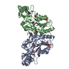 1n2jC  1n2oC S: Starting model for refinement C: citing same article ( |
|---|---|
| Similar structure data | |
| Other databases |

-Links
 PDBj
PDBj


- Assembly
| Deposited unit | 
| ||||||||
|---|---|---|---|---|---|---|---|---|---|
| 1 |
| ||||||||
| Unit cell |
| ||||||||
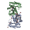
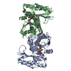
| Details | The biological assembly is a dimer |
-Components
-Protein , 1 types, 2 molecules AB
| #1: Protein | Mass: 31500.100 Da / Num. of mol.: 2 / Mutation: T2A, E77G Source method: isolated from a genetically manipulated source Source: (gene. exp.) Mycobacterium tuberculosis (bacteria) / Gene: panC / Plasmid: pET30a / Species (production host): Escherichia coli / Production host: References: UniProt: P0A5R0, UniProt: P9WIL5*PLUS, pantoate-beta-alanine ligase (AMP-forming) |
|---|
-Non-polymers , 6 types, 320 molecules 


| #2: Chemical |  Mass: 96.063 Da / Num. of mol.: 2 / Source method: obtained synthetically / Formula: SO4 Mass: 96.063 Da / Num. of mol.: 2 / Source method: obtained synthetically / Formula: SO4#3: Chemical |  Mass: 24.305 Da / Num. of mol.: 2 / Source method: obtained synthetically / Formula: Mg Mass: 24.305 Da / Num. of mol.: 2 / Source method: obtained synthetically / Formula: Mg#4: Chemical |  Mass: 505.208 Da / Num. of mol.: 2 / Source method: obtained synthetically / Formula: C11H18N5O12P3 / Comment: AMP-CPP, energy-carrying molecule analogue*YM Mass: 505.208 Da / Num. of mol.: 2 / Source method: obtained synthetically / Formula: C11H18N5O12P3 / Comment: AMP-CPP, energy-carrying molecule analogue*YM#5: Chemical | ChemComp-GOL /  Mass: 92.094 Da / Num. of mol.: 5 / Source method: obtained synthetically / Formula: C3H8O3 Mass: 92.094 Da / Num. of mol.: 5 / Source method: obtained synthetically / Formula: C3H8O3#6: Chemical | ChemComp-EOH /  Mass: 46.068 Da / Num. of mol.: 4 / Source method: obtained synthetically / Formula: C2H6O Mass: 46.068 Da / Num. of mol.: 4 / Source method: obtained synthetically / Formula: C2H6O#7: Water | ChemComp-HOH / | Mass: 18.015 Da / Num. of mol.: 305 / Source method: isolated from a natural source / Formula: H2O |
|---|
-Experimental details
-Experiment
| Experiment | Method: X-RAY DIFFRACTION / Number of used crystals: 1 |
|---|
- Sample preparation
| Crystal | Density Matthews: 2.2 Å3/Da / Density % sol: 43.97 % |
|---|---|
| Crystal grow | Temperature: 293 K / Method: vapor diffusion, hanging drop / pH: 8 Details: PEG3000, lithium sulfate, magnesium sulfate, imidazole, ethanol, glycerol, pH 8.0, VAPOR DIFFUSION, HANGING DROP, temperature 293K |
-Data collection
| Diffraction | Mean temperature: 100 K |
|---|---|
| Diffraction source | Source: ROTATING ANODE / Type: RIGAKU FR-D / Wavelength: 1.5418 |
| Detector | Type: RIGAKU RAXIS IV++ / Detector: IMAGE PLATE / Date: May 4, 2002 / Details: mirrors |
| Radiation | Monochromator: Ni filter / Protocol: SINGLE WAVELENGTH / Monochromatic (M) / Laue (L): M / Scattering type: x-ray |
| Radiation wavelength | Wavelength: 1.5418 Å / Relative weight: 1 |
| Reflection | Resolution: 1.8→50 Å / Num. all: 50403 / Num. obs: 50403 / % possible obs: 99.4 % / Observed criterion σ(I): -3 / Redundancy: 3.4 % / Biso Wilson estimate: 24.4 Å2 / Rmerge(I) obs: 0.059 / Rsym value: 0.059 / Net I/σ(I): 21.1 |
| Reflection shell | Resolution: 1.8→1.86 Å / Redundancy: 3.2 % / Rmerge(I) obs: 0.471 / Mean I/σ(I) obs: 2.6 / Num. unique all: 5035 / Rsym value: 0.471 / % possible all: 99.8 |
- Processing
| Software |
| ||||||||||||||||||||||||||||||||||||||||||||||||||||||||||||||||||||||||||||||||
|---|---|---|---|---|---|---|---|---|---|---|---|---|---|---|---|---|---|---|---|---|---|---|---|---|---|---|---|---|---|---|---|---|---|---|---|---|---|---|---|---|---|---|---|---|---|---|---|---|---|---|---|---|---|---|---|---|---|---|---|---|---|---|---|---|---|---|---|---|---|---|---|---|---|---|---|---|---|---|---|---|---|
| Refinement | Method to determine structure: rigid body refinement into new data Starting model: PDB entry 1MOP Resolution: 1.8→19.84 Å / Rfactor Rfree error: 0.003 / Isotropic thermal model: RESTRAINED / Cross valid method: THROUGHOUT / σ(F): 0 / Stereochemistry target values: Engh & Huber
| ||||||||||||||||||||||||||||||||||||||||||||||||||||||||||||||||||||||||||||||||
| Solvent computation | Solvent model: FLAT MODEL / Bsol: 49.0266 Å2 / ksol: 0.370081 e/Å3 | ||||||||||||||||||||||||||||||||||||||||||||||||||||||||||||||||||||||||||||||||
| Displacement parameters | Biso mean: 30 Å2
| ||||||||||||||||||||||||||||||||||||||||||||||||||||||||||||||||||||||||||||||||
| Refine analyze |
| ||||||||||||||||||||||||||||||||||||||||||||||||||||||||||||||||||||||||||||||||
| Refinement step | Cycle: LAST / Resolution: 1.8→19.84 Å
| ||||||||||||||||||||||||||||||||||||||||||||||||||||||||||||||||||||||||||||||||
| Refine LS restraints |
| ||||||||||||||||||||||||||||||||||||||||||||||||||||||||||||||||||||||||||||||||
| Refine LS restraints NCS | NCS model details: RESTRAIN | ||||||||||||||||||||||||||||||||||||||||||||||||||||||||||||||||||||||||||||||||
| LS refinement shell | Resolution: 1.8→1.91 Å / Rfactor Rfree error: 0.011 / Total num. of bins used: 6
| ||||||||||||||||||||||||||||||||||||||||||||||||||||||||||||||||||||||||||||||||
| Xplor file |
|
